Coloured TEM of pancreatic acinar secretory cells
Bildnummer 11872976
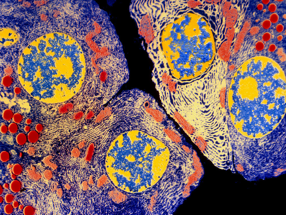
| Pancreatic acinar cell. Coloured transmission electron micrograph (TEM) of a slice through several enzyme-secreting acinar cells from a pancreas. The red circles are secretory granules packed with digestive enzymes ready for export to the small intestine. The four round blue and yellow structures are cell nuclei. Each cell is filled with a network of densely folded membrane,called rough endoplasmic reticulum,here coloured light yellow. The membrane's surface is covered in small dots,called ribosomes. These are protein-manufacturing sites where various digestive enzymes are produced and secreted by the cell. Magnification x1200 at 6x4.5cm size | |
| Lizenzart: | Lizenzpflichtig |
| Credit: | Science Photo Library |
| Bildgröße: | 4501 px × 3385 px |
| Modell-Rechte: | nicht erforderlich |
| Eigentums-Rechte: | nicht erforderlich |
| Restrictions: | - |
Preise für dieses Bild ab 15 €
Universitäten & Organisationen
(Informationsmaterial Digital, Informationsmaterial Print, Lehrmaterial Digital etc.)
ab 15 €
Redaktionell
(Bücher, Bücher: Sach- und Fachliteratur, Digitale Medien (redaktionell) etc.)
ab 30 €
Werbung
(Anzeigen, Aussenwerbung, Digitale Medien, Fernsehwerbung, Karten, Werbemittel, Zeitschriften etc.)
ab 55 €
Handelsprodukte
(bedruckte Textilie, Kalender, Postkarte, Grußkarte, Verpackung etc.)
ab 75 €
Pauschalpreise
Rechtepakete für die unbeschränkte Bildnutzung in Print oder Online
ab 495 €