Gall bladder surface,SEM
Bildnummer 11872929
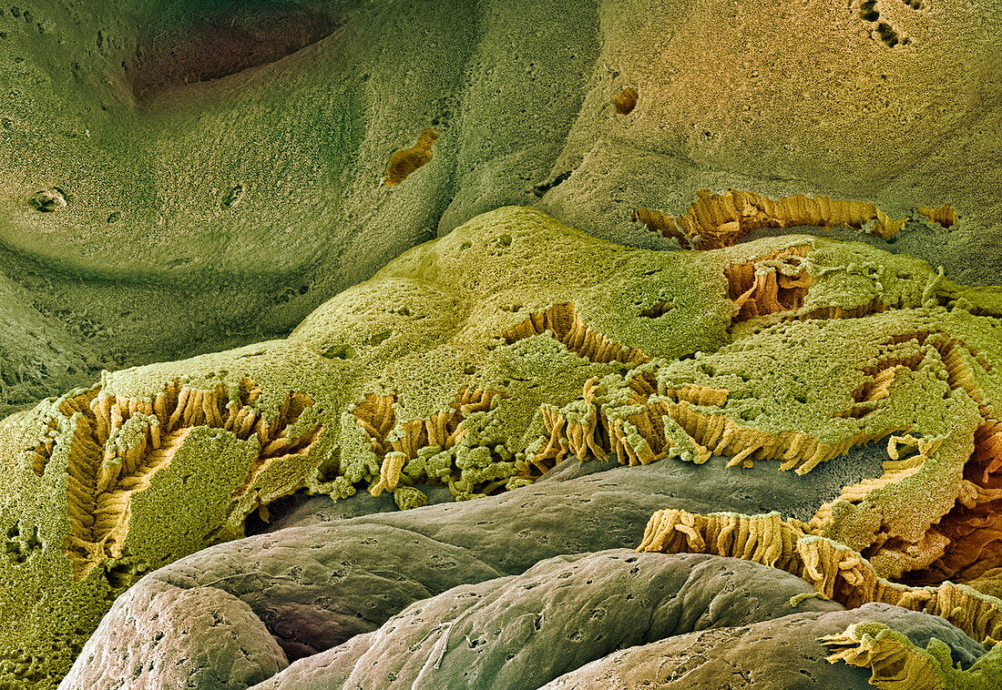
| Gall bladder. Coloured scanning electron micrograph (SEM) of the internal surface of a gall bladder. This mucosa lining is made up of columnar epithelial cells (green and yellow). Each cell of the gall bladder lining has microvilli (tiny projections) that increase its surface area and aid water uptake. Connective tissue (brown) is seen below the lining where some of the cells have come away. The gall bladder is a sac that concentrates and stores bile,produced by the liver,and releases it into the duodenum (small intestine),where it aids the digestion of fats. Magnification: x55 when printed 10 centimetres wide | |
| Lizenzart: | Lizenzpflichtig |
| Credit: | Science Photo Library / Gschmeissner, Steve |
| Bildgröße: | 3500 px × 2411 px |
| Modell-Rechte: | nicht erforderlich |
| Eigentums-Rechte: | nicht erforderlich |
| Restrictions: | - |
Preise für dieses Bild ab 15 €
Universitäten & Organisationen
(Informationsmaterial Digital, Informationsmaterial Print, Lehrmaterial Digital etc.)
ab 15 €
Redaktionell
(Bücher, Bücher: Sach- und Fachliteratur, Digitale Medien (redaktionell) etc.)
ab 30 €
Werbung
(Anzeigen, Aussenwerbung, Digitale Medien, Fernsehwerbung, Karten, Werbemittel, Zeitschriften etc.)
ab 55 €
Handelsprodukte
(bedruckte Textilie, Kalender, Postkarte, Grußkarte, Verpackung etc.)
ab 75 €
Pauschalpreise
Rechtepakete für die unbeschränkte Bildnutzung in Print oder Online
ab 495 €
Keywords
- Anatomie,
- Bindegewebe,
- Biologie,
- biologisch,
- eingefärbt,
- epithelial,
- farbig,
- Galle,
- gefärbt,
- Histologie,
- histologisch,
- Lager,
- menschlicher Körper,
- Mikrovilli,
- Mikrovillus,
- Oberfläche,
- Organ,
- Rasterelektronenmikroskop,
- rasterelektronenmikroskopische Aufnahme,
- REM,
- Schleimhaut,
- Verdauung,
- Verdauungs-,
- Zelle,
- Zellen