False-colour SEM of surface peritoneum of liver
Bildnummer 11872875
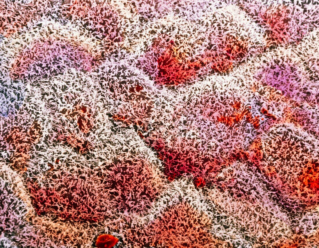
| False-colour scanning electron micrograph (SEM) of the surface peritoneal layer of cells around the liver. Covered with many short microvilli (hair- like structures),these flattened serosal cells form an organ holding sac around the liver. Its surface area,increased by the microvilli,secre- tes fluid derived from blood serum: this allows a frictionless movement of the liver within the abdomen. Such a protective layer for organs is important,especially in the abdomen,where many organs coexist and function during body movement. Beneath microvilli the outline of individual cells can be seen. Magnification: x1,125 at 6x7cm size. Magnification: x1,800 at 4x5 inch size | |
| Lizenzart: | Lizenzpflichtig |
| Credit: | Science Photo Library / UNIVERSITY LA SAPIENZA, ROME / DEPT. OF ANATOMY / PROF. P. MOTTA |
| Bildgröße: | 4843 px × 3779 px |
| Modell-Rechte: | nicht erforderlich |
| Eigentums-Rechte: | nicht erforderlich |
| Restrictions: | - |
Preise für dieses Bild ab 15 €
Universitäten & Organisationen
(Informationsmaterial Digital, Informationsmaterial Print, Lehrmaterial Digital etc.)
ab 15 €
Redaktionell
(Bücher, Bücher: Sach- und Fachliteratur, Digitale Medien (redaktionell) etc.)
ab 30 €
Werbung
(Anzeigen, Aussenwerbung, Digitale Medien, Fernsehwerbung, Karten, Werbemittel, Zeitschriften etc.)
ab 55 €
Handelsprodukte
(bedruckte Textilie, Kalender, Postkarte, Grußkarte, Verpackung etc.)
ab 75 €
Pauschalpreise
Rechtepakete für die unbeschränkte Bildnutzung in Print oder Online
ab 495 €