Small intestine villus
Bildnummer 11872824
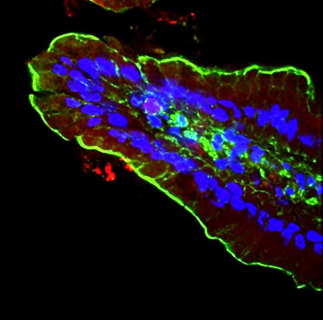
| Small intestine villus,fluorescence deconvolution micrograph. Fluorescent dyes have been used to highlight cellular structures and proteins: microvilli actin (green),cell nuclei (blue). Villi are the functional structures lining the walls of the intestines. Actin is a protein that is a major part of a cell's cytoskeleton,here highlighted in the microvilli. These are small structures,covering the surface of a villus,which absorb nutrients from digested food. The cell nuclei seen here are part of the columnar epithelium that forms the shape of a villus | |
| Lizenzart: | Lizenzpflichtig |
| Credit: | Science Photo Library / R. BICK, B. POINDEXTER, UT MEDICAL SCHOOL |
| Bildgröße: | 3300 px × 3280 px |
| Modell-Rechte: | nicht erforderlich |
| Eigentums-Rechte: | nicht erforderlich |
| Restrictions: | - |
Preise für dieses Bild ab 15 €
Universitäten & Organisationen
(Informationsmaterial Digital, Informationsmaterial Print, Lehrmaterial Digital etc.)
ab 15 €
Redaktionell
(Bücher, Bücher: Sach- und Fachliteratur, Digitale Medien (redaktionell) etc.)
ab 30 €
Werbung
(Anzeigen, Aussenwerbung, Digitale Medien, Fernsehwerbung, Karten, Werbemittel, Zeitschriften etc.)
ab 55 €
Handelsprodukte
(bedruckte Textilie, Kalender, Postkarte, Grußkarte, Verpackung etc.)
ab 75 €
Pauschalpreise
Rechtepakete für die unbeschränkte Bildnutzung in Print oder Online
ab 495 €
Keywords
- Aktin,
- Anatomie,
- anatomisch,
- Biochemie,
- biochemisch,
- dapi,
- Darm,
- Darm-,
- Dünndarm,
- einer,
- Eiweiß,
- Farbstoff,
- Fluoreszenz,
- Fluoreszenzentfaltung,
- fluoreszierend,
- Gewebe,
- Histologie,
- histologisch,
- Lichtmikroskop,
- Marker,
- menschlicher Körper,
- Mikrofotografie,
- Mikrovillus,
- Single,
- Verdauung,
- Verdauungssystem,
- Zellbilogie,
- Zelle,
- Zellen,
- Zellkern,
- Zotte,
- Zytologie