Intestinal lining,SEM
Bildnummer 11872811
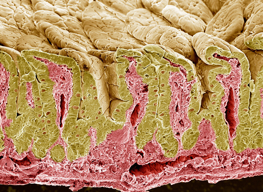
| Intestinal lining. Coloured scanning electron micrograph (SEM) of a fractured intestinal surface. The fracture plane (lower frame) has cut down through the surface,revealing the deep folds called villi. The intestinal surface (brown) is where food is digested. It consists of a layer of surface (epithelial) cells (yellow),with goblet cells (small circular cells,pink) that secrete mucus to lubricate food and prevent self-digestion. This outer surface is supported by connective tissue (pink),seen at the core of each fold (villus). The folds increase the area for the absorption of food. Magnification: x200 when printed 10 centimetres wide | |
| Lizenzart: | Lizenzpflichtig |
| Credit: | Science Photo Library / Gschmeissner, Steve |
| Bildgröße: | 4100 px × 2993 px |
| Modell-Rechte: | nicht erforderlich |
| Eigentums-Rechte: | nicht erforderlich |
| Restrictions: | - |
Preise für dieses Bild ab 15 €
Universitäten & Organisationen
(Informationsmaterial Digital, Informationsmaterial Print, Lehrmaterial Digital etc.)
ab 15 €
Redaktionell
(Bücher, Bücher: Sach- und Fachliteratur, Digitale Medien (redaktionell) etc.)
ab 30 €
Werbung
(Anzeigen, Aussenwerbung, Digitale Medien, Fernsehwerbung, Karten, Werbemittel, Zeitschriften etc.)
ab 55 €
Handelsprodukte
(bedruckte Textilie, Kalender, Postkarte, Grußkarte, Verpackung etc.)
ab 75 €
Pauschalpreise
Rechtepakete für die unbeschränkte Bildnutzung in Print oder Online
ab 495 €
Keywords
- Anatomie,
- anatomisch,
- Biologie,
- biologisch,
- Darm,
- Darm-,
- Dünndarm,
- Einfrieren,
- Epithel,
- epithelial,
- Falten,
- farbig,
- frakturiert,
- Futter,
- Gefaltet,
- gefärbt,
- gefriergebrochen,
- gesund,
- Gewebe,
- Histologie,
- histologisch,
- Kelch,
- Mauer,
- menschlicher Körper,
- normal,
- Oberfläche,
- Rasterelektronenmikroskop,
- rasterelektronenmikroskopische Aufnahme,
- REM,
- sekretorisch,
- Sektion,
- sektioniert,
- Verdauung,
- Verdauungssystem,
- Zelle,
- Zellen,
- Zotte,
- Zotten