Small intestine
Bildnummer 11872786
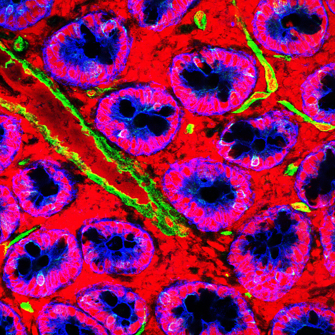
| Small intestine. Fluorescence confocal light micrograph of a horizontal section through the mucosa of the human small intestine,showing crypts of Lieberkuhn (pink and blue). The small intestine runs from the stomach to the large intestine. It is where digestion is completed and nutrients are absorbed into the blood. The crypts secrete enzymes into the interior (lumen) of the intestine that help to digest the food. Cell nuclei have been stained with propidium iodide (red) and epithelial cells have been stained with a cytokeratin-specific antibody (blue). Shiga toxin (Stx,green),which was added after the sections were cut,is stained with an Stx-specific antibody. In this section Stx has bound to small blood vessels | |
| Lizenzart: | Lizenzpflichtig |
| Credit: | Science Photo Library / Schuller, Stephanie |
| Bildgröße: | 2958 px × 2958 px |
| Modell-Rechte: | nicht erforderlich |
| Eigentums-Rechte: | nicht erforderlich |
| Restrictions: | - |
Preise für dieses Bild ab 15 €
Universitäten & Organisationen
(Informationsmaterial Digital, Informationsmaterial Print, Lehrmaterial Digital etc.)
ab 15 €
Redaktionell
(Bücher, Bücher: Sach- und Fachliteratur, Digitale Medien (redaktionell) etc.)
ab 30 €
Werbung
(Anzeigen, Aussenwerbung, Digitale Medien, Fernsehwerbung, Karten, Werbemittel, Zeitschriften etc.)
ab 55 €
Handelsprodukte
(bedruckte Textilie, Kalender, Postkarte, Grußkarte, Verpackung etc.)
ab 75 €
Pauschalpreise
Rechtepakete für die unbeschränkte Bildnutzung in Print oder Online
ab 495 €
Keywords
- Anatomie,
- anatomisch,
- befleckt,
- Biologie,
- biologisch,
- Darm,
- Dünndarm,
- Fluoreszenz,
- Fluoreszenzlichtmikroskopische Aufnahme,
- fluoreszierend,
- gesund,
- Histologie,
- histologisch,
- konfokales Lichtmikroskop,
- Lichtmikroskop,
- lichtmikroskopische Aufnahme,
- menschlicher Körper,
- normal,
- Sektion,
- sektioniert,
- Verdauung,
- Verdauungssystem,
- Verfärbung,
- Zelle,
- Zellen,
- Zotte,
- Zotten