Intestinal villi
Bildnummer 11872743
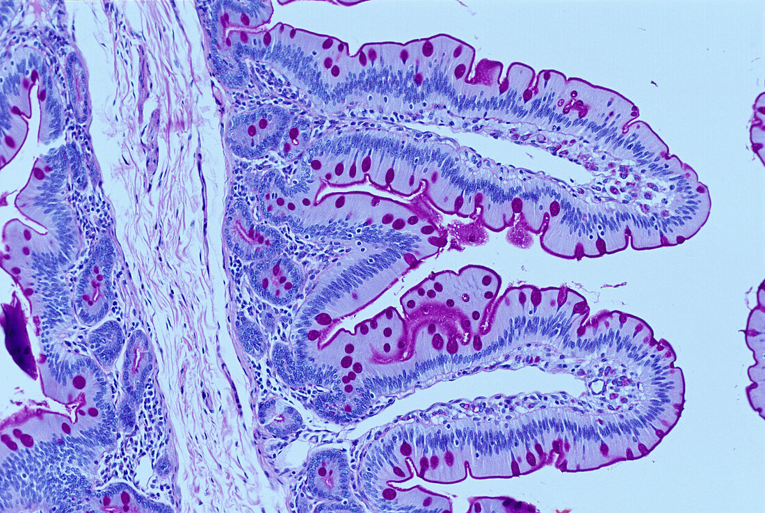
| Intestinal villi. Light micrograph of villi on the lining of the small intestine. Each fold of the wall of the small intestine is covered with these small projections called villi,which serve to increase the surface area for the absorption of nutrients from the gut lumen. Villi are mainly composed of columnar epithelial cells (purple) interspersed with goblet cells (pink). The goblet cells secrete mucous,which lines the surface of the membrane. The regularly ordered nuclei of the columnar cells are easily visible. The empty core of each villus is a lymphatic vessel that removes cellular waste. Magnification: x115 when printed 10cm wide | |
| Lizenzart: | Lizenzpflichtig |
| Credit: | Science Photo Library / Innerspace Imaging |
| Bildgröße: | 5120 px × 3429 px |
| Modell-Rechte: | nicht erforderlich |
| Eigentums-Rechte: | nicht erforderlich |
| Restrictions: | - |
Preise für dieses Bild ab 15 €
Universitäten & Organisationen
(Informationsmaterial Digital, Informationsmaterial Print, Lehrmaterial Digital etc.)
ab 15 €
Redaktionell
(Bücher, Bücher: Sach- und Fachliteratur, Digitale Medien (redaktionell) etc.)
ab 30 €
Werbung
(Anzeigen, Aussenwerbung, Digitale Medien, Fernsehwerbung, Karten, Werbemittel, Zeitschriften etc.)
ab 55 €
Handelsprodukte
(bedruckte Textilie, Kalender, Postkarte, Grußkarte, Verpackung etc.)
ab 75 €
Pauschalpreise
Rechtepakete für die unbeschränkte Bildnutzung in Print oder Online
ab 495 €
Keywords
- Absorption,
- Anatomie,
- anatomisch,
- befleckt,
- Biologie,
- biologisch,
- Darm,
- eingefärbt,
- Enterozyten,
- Epithel,
- Falten,
- farbig,
- Futter,
- Gefaltet,
- gefärbt,
- gesund,
- Histologie,
- histologisch,
- Jejunum,
- klein,
- kreisförmig,
- Lichtmikroskop,
- lichtmikroskopische Aufnahme,
- Lymphgang,
- Membran,
- Mensch,
- menschlicher Körper,
- Mikroplicae,
- normal,
- Physiologie,
- physiologisch,
- Schleimhaut,
- Sektion,
- sektioniert,
- Struktur,
- Verdauung,
- Verdauungskanal,
- Verdauungstrakt,
- Zotte,
- Zotten,
- Zytologie,
- Zytologisch