Small intestine lining
Bildnummer 11872720
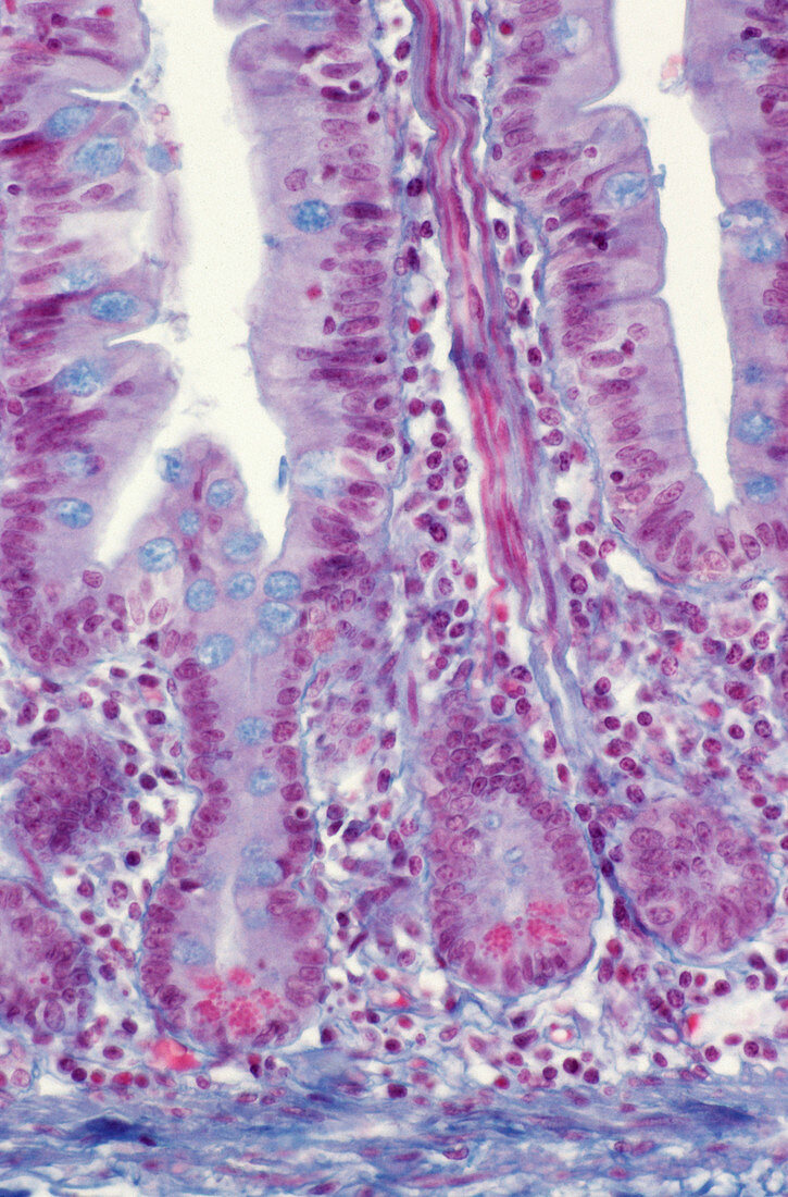
| Small intestine lining. Light micrograph of a section through the lining (mucosa) of the small intestine. It shows crypts of Lieberkuhn (purple folds,lower) between the intestinal villi (projections,upper). The crypts produce new cells by constant cell division; these progress up the villi to renew the intestinal lining. The lining of the villi and crypts is composed of columnar enterocyte cells,which are involved in digestion and absorption,and mucus-secreting goblet cells (blue). At the base of the crypts are Paneth cells (stained red),which are thought to protect the intestine from disease. At bottom is a muscle layer (blue). Magnification unknown | |
| Lizenzart: | Lizenzpflichtig |
| Credit: | Science Photo Library / CNRI |
| Bildgröße: | 3045 px × 4621 px |
| Modell-Rechte: | nicht erforderlich |
| Eigentums-Rechte: | nicht erforderlich |
| Restrictions: | - |
Preise für dieses Bild ab 15 €
Universitäten & Organisationen
(Informationsmaterial Digital, Informationsmaterial Print, Lehrmaterial Digital etc.)
ab 15 €
Redaktionell
(Bücher, Bücher: Sach- und Fachliteratur, Digitale Medien (redaktionell) etc.)
ab 30 €
Werbung
(Anzeigen, Aussenwerbung, Digitale Medien, Fernsehwerbung, Karten, Werbemittel, Zeitschriften etc.)
ab 55 €
Handelsprodukte
(bedruckte Textilie, Kalender, Postkarte, Grußkarte, Verpackung etc.)
ab 75 €
Pauschalpreise
Rechtepakete für die unbeschränkte Bildnutzung in Print oder Online
ab 495 €
Keywords
- Anatomie,
- befleckt,
- Darm,
- Dünndarm,
- Einteilung,
- Enterozyten,
- Futter,
- gesund,
- Histologie,
- histologisch,
- Ileum,
- kapillar,
- Krypta,
- Krypten von Lieberkuhn,
- lichtmikroskopische Aufnahme,
- menschlicher Körper,
- Mitose,
- normal,
- rutschen,
- Schleimhaut,
- Sektion,
- sektioniert,
- Trakt,
- Verdauung,
- Verdauungskanal,
- Verdauungssystem,
- Verfärbung,
- Zellen