Small intestine villus,SEM
Bildnummer 11872711
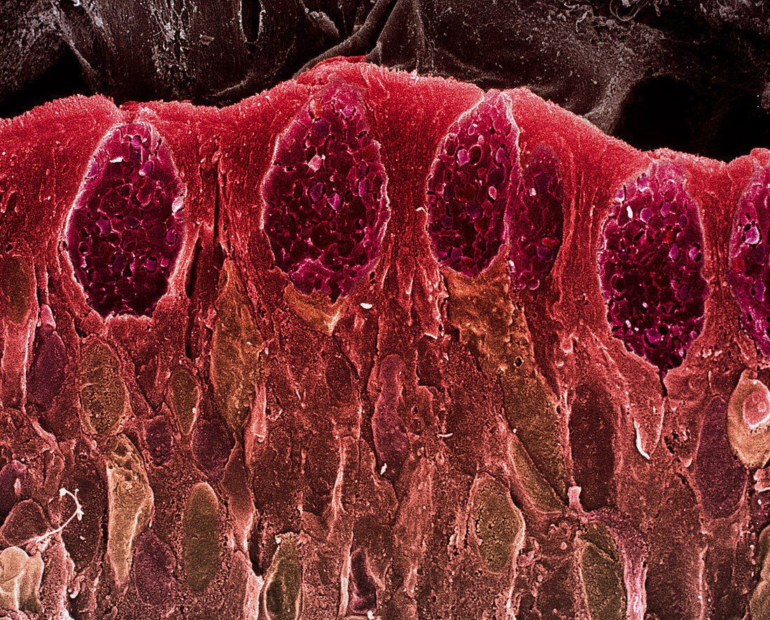
| Small intestine villus. Coloured scanning electron micrograph (SEM) of a freeze fracture section through a villus from the mucosal lining of the small intestine. Villi are finger-like projections that increase the surface area of a structure. Microvilli,just visible across upper centre,further increase the surface area available for food absorption. The outer surface of a villus is mostly columnar epithelium (red). It contains numerous goblet cells (dark pink),which secrete mucus to lubricate food & prevent self-digestion. Within the goblet cells individual mucin granules are seen. Magnification: x2000 at 6x7cm size | |
| Lizenzart: | Lizenzpflichtig |
| Credit: | Science Photo Library / Gschmeissner, Steve |
| Bildgröße: | 5298 px × 4266 px |
| Modell-Rechte: | nicht erforderlich |
| Eigentums-Rechte: | nicht erforderlich |
| Restrictions: | - |
Preise für dieses Bild ab 15 €
Universitäten & Organisationen
(Informationsmaterial Digital, Informationsmaterial Print, Lehrmaterial Digital etc.)
ab 15 €
Redaktionell
(Bücher, Bücher: Sach- und Fachliteratur, Digitale Medien (redaktionell) etc.)
ab 30 €
Werbung
(Anzeigen, Aussenwerbung, Digitale Medien, Fernsehwerbung, Karten, Werbemittel, Zeitschriften etc.)
ab 55 €
Handelsprodukte
(bedruckte Textilie, Kalender, Postkarte, Grußkarte, Verpackung etc.)
ab 75 €
Pauschalpreise
Rechtepakete für die unbeschränkte Bildnutzung in Print oder Online
ab 495 €
Keywords
- Absorption,
- Anatomie,
- Darm,
- Darm-,
- Dünndarm,
- Enterozyten,
- Epithel,
- epithelial,
- farbig,
- frakturiert,
- Futter,
- gesund,
- Gewebe,
- Histologie,
- Membran,
- menschlicher Körper,
- Mikrovilli,
- normal,
- Oberfläche,
- rasterelektronenmikroskopische Aufnahme,
- REM,
- Schleim,
- Schleimhaut,
- sekretorisch,
- Verdauung,
- Verdauungskanal,
- Verdauungssystem,
- Zellen,
- Zotte,
- Zotten