Small intestine mucosa
Bildnummer 11872708
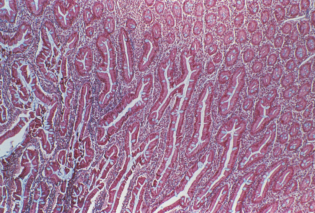
| Small intestine lining. Light micrograph of a section through the lining (mucosa) of the ileum,part of the small intestine,showing the numerous crypts of Lieberkuhn (purple/white). The crypts are seen in both transverse section (upper left to bottom right) and in longitudinal section (rest of frame). They lie between the intestinal villi (finger-like projections of the lining,not seen),producing new cells which mature and replace dead cells lost into the lumen (cavity) of the intestine. The crypts are composed of columnar enterocyte cells (purple) and mucus-secreting goblet cells (light blue). Trichrome blue stain. Magnification unknown | |
| Lizenzart: | Lizenzpflichtig |
| Credit: | Science Photo Library / CNRI |
| Bildgröße: | 4575 px × 3096 px |
| Modell-Rechte: | nicht erforderlich |
| Eigentums-Rechte: | nicht erforderlich |
| Restrictions: | - |
Preise für dieses Bild ab 15 €
Universitäten & Organisationen
(Informationsmaterial Digital, Informationsmaterial Print, Lehrmaterial Digital etc.)
ab 15 €
Redaktionell
(Bücher, Bücher: Sach- und Fachliteratur, Digitale Medien (redaktionell) etc.)
ab 30 €
Werbung
(Anzeigen, Aussenwerbung, Digitale Medien, Fernsehwerbung, Karten, Werbemittel, Zeitschriften etc.)
ab 55 €
Handelsprodukte
(bedruckte Textilie, Kalender, Postkarte, Grußkarte, Verpackung etc.)
ab 75 €
Pauschalpreise
Rechtepakete für die unbeschränkte Bildnutzung in Print oder Online
ab 495 €