False-colour SEM of zone between ileum and caecum
Bildnummer 11872665
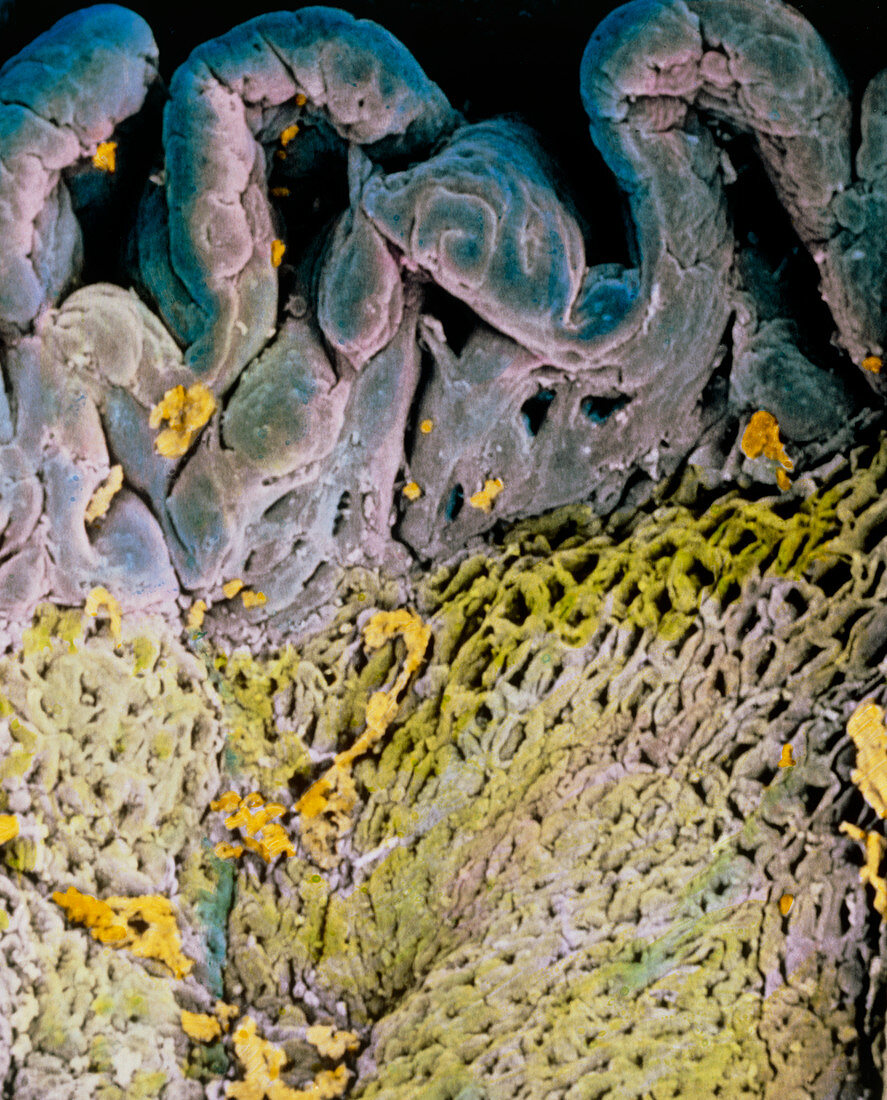
| False-colour scanning electron micrograph (SEM) of the transition zone between the small intestine (ileum,blue) and large intestine (caecum,green). At above centre,the ileum wall is pleated with plicae and smaller finger-like villi. These folds increase the surface area of the small intestine for its role in food digestion and absorption. Digested food is absorbed into the blood through capillaries in each villus. By contrast the caecum wall is flat,with many openings to mucous glands. In the large intestine,faeces form by absorption of excess water while mucous lubricates their path. Magnification: x100 at 6x7cm size. Magnification: x160 at 4x5 inch size | |
| Lizenzart: | Lizenzpflichtig |
| Credit: | Science Photo Library / UNIVERSITY LA SAPIENZA, ROME / DEPT. OF ANATOMY / PROF. P. MOTTA |
| Bildgröße: | 4029 px × 4994 px |
| Modell-Rechte: | nicht erforderlich |
| Eigentums-Rechte: | nicht erforderlich |
| Restrictions: | - |
Preise für dieses Bild ab 15 €
Universitäten & Organisationen
(Informationsmaterial Digital, Informationsmaterial Print, Lehrmaterial Digital etc.)
ab 15 €
Redaktionell
(Bücher, Bücher: Sach- und Fachliteratur, Digitale Medien (redaktionell) etc.)
ab 30 €
Werbung
(Anzeigen, Aussenwerbung, Digitale Medien, Fernsehwerbung, Karten, Werbemittel, Zeitschriften etc.)
ab 55 €
Handelsprodukte
(bedruckte Textilie, Kalender, Postkarte, Grußkarte, Verpackung etc.)
ab 75 €
Pauschalpreise
Rechtepakete für die unbeschränkte Bildnutzung in Print oder Online
ab 495 €