F/colour SEM of oesophagus-stomach transition zone
Bildnummer 11872553
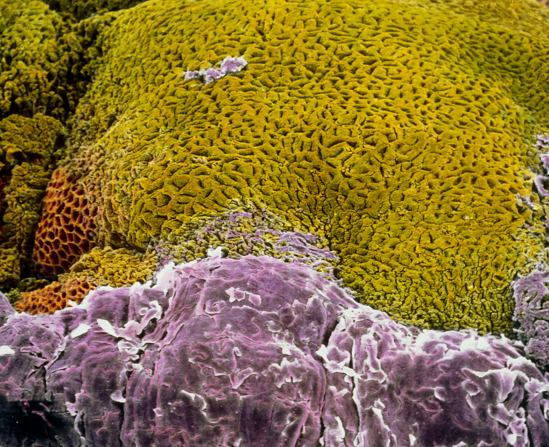
| False-colour scanning electron micrograph (SEM) of the transition zone between the oesophagus and stomach. Below,the stratified squamous epithelium of the oesophagus (violet),contrasts with the glandular wall of the stomach mucosa (green). The surface of the oesophagus is thick,multi-layered,and adapted for the movement of food along it. While the stomach mucosa shows many orifices of glands which secrete digestive enzymes,hydrochloric acid,and hormones; a simple columnar epithelium secretes mucous. At left (brown),part of the stomach wall is destroyed in an ulcer-like manner. Magnification: x90 at 6x7cm size. Magnification: x140 at 4x5 inch size | |
| Lizenzart: | Lizenzpflichtig |
| Credit: | Science Photo Library / UNIVERSITY LA SAPIENZA, ROME / DEPT. OF ANATOMY / PROF. P. MOTTA |
| Bildgröße: | 4022 px × 3277 px |
| Modell-Rechte: | nicht erforderlich |
| Eigentums-Rechte: | nicht erforderlich |
| Restrictions: | - |
Preise für dieses Bild ab 15 €
Universitäten & Organisationen
(Informationsmaterial Digital, Informationsmaterial Print, Lehrmaterial Digital etc.)
ab 15 €
Redaktionell
(Bücher, Bücher: Sach- und Fachliteratur, Digitale Medien (redaktionell) etc.)
ab 30 €
Werbung
(Anzeigen, Aussenwerbung, Digitale Medien, Fernsehwerbung, Karten, Werbemittel, Zeitschriften etc.)
ab 55 €
Handelsprodukte
(bedruckte Textilie, Kalender, Postkarte, Grußkarte, Verpackung etc.)
ab 75 €
Pauschalpreise
Rechtepakete für die unbeschränkte Bildnutzung in Print oder Online
ab 495 €