Coloured SEM of filiform papillae on the tongue
Bildnummer 11872342
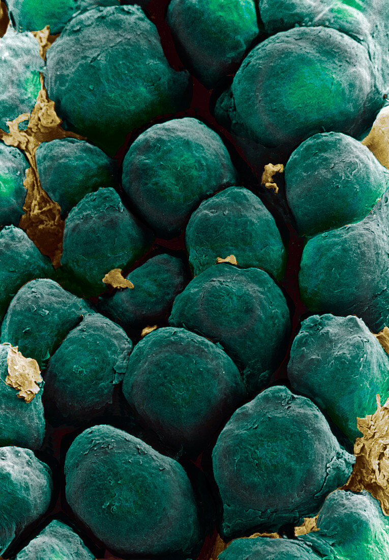
| Tongue papillae. Coloured scanning electron micrograph (SEM) of filiform papillae on the surface of the tongue. Filiform papillae are covered by stratified squamous epithelial cells. Dead cells of the uppermost layer are constantly being shed and replaced (desquamation). This shedding gives the papillae their scaly appearance. Filiform papillae form a rough surface to aid chewing. Each papilla contains nerve endings which transmit tactile (touch) information to the brain. Magnification unknown | |
| Lizenzart: | Lizenzpflichtig |
| Credit: | Science Photo Library / Gschmeissner, Steve |
| Bildgröße: | 2542 px × 3655 px |
| Modell-Rechte: | nicht erforderlich |
| Eigentums-Rechte: | nicht erforderlich |
| Restrictions: | - |
Preise für dieses Bild ab 15 €
Universitäten & Organisationen
(Informationsmaterial Digital, Informationsmaterial Print, Lehrmaterial Digital etc.)
ab 15 €
Redaktionell
(Bücher, Bücher: Sach- und Fachliteratur, Digitale Medien (redaktionell) etc.)
ab 30 €
Werbung
(Anzeigen, Aussenwerbung, Digitale Medien, Fernsehwerbung, Karten, Werbemittel, Zeitschriften etc.)
ab 55 €
Handelsprodukte
(bedruckte Textilie, Kalender, Postkarte, Grußkarte, Verpackung etc.)
ab 75 €
Pauschalpreise
Rechtepakete für die unbeschränkte Bildnutzung in Print oder Online
ab 495 €