Cochlea cells,SEM
Bildnummer 11872147
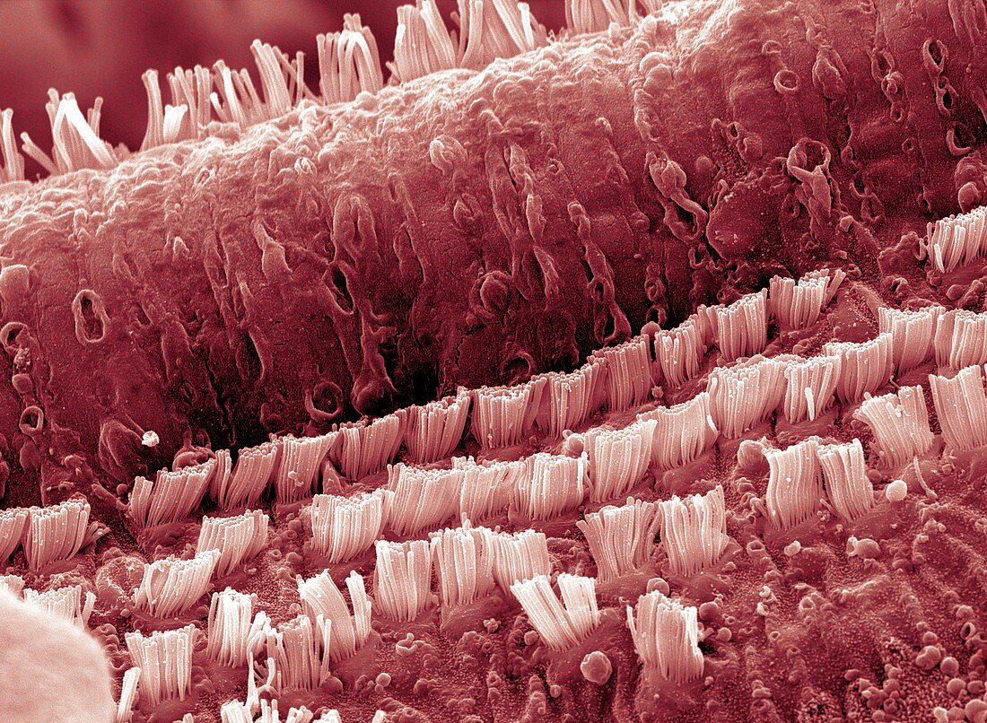
| Cochlea cells. Coloured scanning electron micrograph (SEM) of hair cells in the cochlea of a human ear. The rows of columnar outer pillar cells run along the organ of Corti,the auditory sense organ. The outer pillar cells arise from the basilar membrane,and their upper surfaces form part of the surface of the organ of Corti. This organ lies on the basilar membrane,an internal surface of the cochlear duct. The tectorial membrane,which overlies the sensory hairs,has been removed. Sound waves deform hair cell cilia & trigger auditory nerve impulses. Magnification: x5000 at 6x7cm size | |
| Lizenzart: | Lizenzpflichtig |
| Credit: | Science Photo Library / Clouds Hill Imaging |
| Bildgröße: | 4096 px × 3002 px |
| Modell-Rechte: | nicht erforderlich |
| Eigentums-Rechte: | nicht erforderlich |
| Restrictions: | - |
Preise für dieses Bild ab 15 €
Universitäten & Organisationen
(Informationsmaterial Digital, Informationsmaterial Print, Lehrmaterial Digital etc.)
ab 15 €
Redaktionell
(Bücher, Bücher: Sach- und Fachliteratur, Digitale Medien (redaktionell) etc.)
ab 30 €
Werbung
(Anzeigen, Aussenwerbung, Digitale Medien, Fernsehwerbung, Karten, Werbemittel, Zeitschriften etc.)
ab 55 €
Handelsprodukte
(bedruckte Textilie, Kalender, Postkarte, Grußkarte, Verpackung etc.)
ab 75 €
Pauschalpreise
Rechtepakete für die unbeschränkte Bildnutzung in Print oder Online
ab 495 €