SEM of hair cells
Bildnummer 11872137
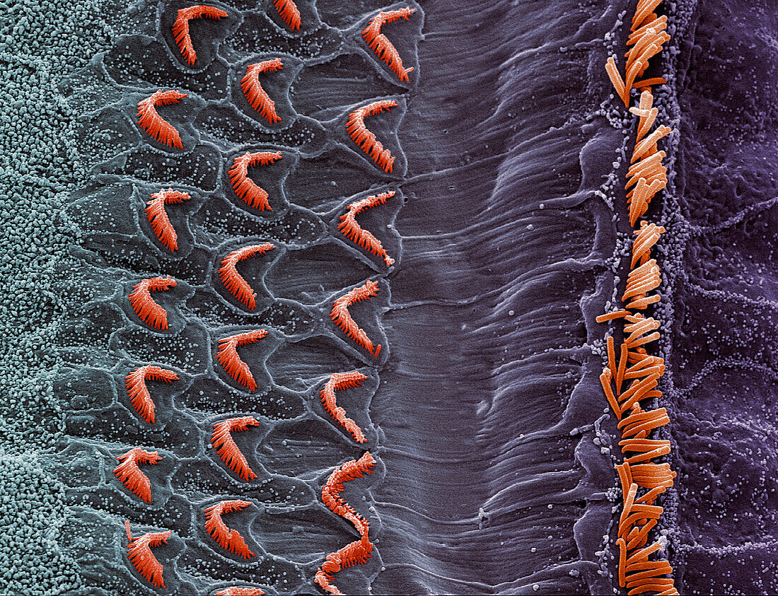
| Inner ear hair cells. Coloured scanning electron micrograph (SEM) of hair cells in the organ of Corti. This delicate structure in the cochlea of the inner ear converts sound vibrations into nerve impulses. The three rows of outer hair cells have V-shaped groups of stereocilia (red),while the single row of inner hair cells is shown by a line of stereocilia. The hair cells sit between two membranes (neither seen) in endolymph fluid. Sound vibrations transmitted from the perilymph fluid of the middle ear into the endolymph cause the hair cells to compress,generating nerve impulses that are carried to the brain. Magnification unknown | |
| Lizenzart: | Lizenzpflichtig |
| Credit: | Science Photo Library / Gschmeissner, Steve |
| Bildgröße: | 2717 px × 2083 px |
| Modell-Rechte: | nicht erforderlich |
| Eigentums-Rechte: | nicht erforderlich |
| Restrictions: | - |
Preise für dieses Bild ab 15 €
Universitäten & Organisationen
(Informationsmaterial Digital, Informationsmaterial Print, Lehrmaterial Digital etc.)
ab 15 €
Redaktionell
(Bücher, Bücher: Sach- und Fachliteratur, Digitale Medien (redaktionell) etc.)
ab 30 €
Werbung
(Anzeigen, Aussenwerbung, Digitale Medien, Fernsehwerbung, Karten, Werbemittel, Zeitschriften etc.)
ab 55 €
Handelsprodukte
(bedruckte Textilie, Kalender, Postkarte, Grußkarte, Verpackung etc.)
ab 75 €
Pauschalpreise
Rechtepakete für die unbeschränkte Bildnutzung in Print oder Online
ab 495 €