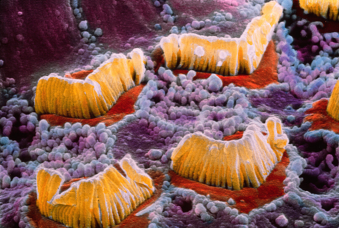
SEM of hair cells in organ of Corti
Bildnummer 11872121

| False-colour scanning electron micrograph of hair cells (yellow v-shaped structures) which are part of the organ of Corti in the inner ear. This organ is situated inside the cochlea and converts the mechanical energy of sound waves into electrical stimuli. The hair cells are immersed in a fluid called endolymph and react to the pressure of sound waves with an undulatory motion. This motion stimulates a nerve ending in each group of hair cells which carries a signal to various parts of the brain through the Cochlear nerve. Epithelial cells are covered here by microvilli (purple spheres). Magnification: x3900 at 6x7cm size | |
| Lizenzart: | Lizenzpflichtig |
| Credit: | Science Photo Library / UNIVERSITY LA SAPIENZA, ROME / DEPT. OF ANATOMY / PROF. P. MOTTA |
| Bildgröße: | 4567 px × 3075 px |
| Modell-Rechte: | nicht erforderlich |
| Eigentums-Rechte: | nicht erforderlich |
| Restrictions: | - |
Preise für dieses Bild ab 15 €
Universitäten & Organisationen
(Informationsmaterial Digital, Informationsmaterial Print, Lehrmaterial Digital etc.)
ab 15 €
Redaktionell
(Bücher, Bücher: Sach- und Fachliteratur, Digitale Medien (redaktionell) etc.)
ab 30 €
Werbung
(Anzeigen, Aussenwerbung, Digitale Medien, Fernsehwerbung, Karten, Werbemittel, Zeitschriften etc.)
ab 55 €
Handelsprodukte
(bedruckte Textilie, Kalender, Postkarte, Grußkarte, Verpackung etc.)
ab 75 €
Pauschalpreise
Rechtepakete für die unbeschränkte Bildnutzung in Print oder Online
ab 495 €
