Retina blood vessel and nerve cells
Bildnummer 11872007
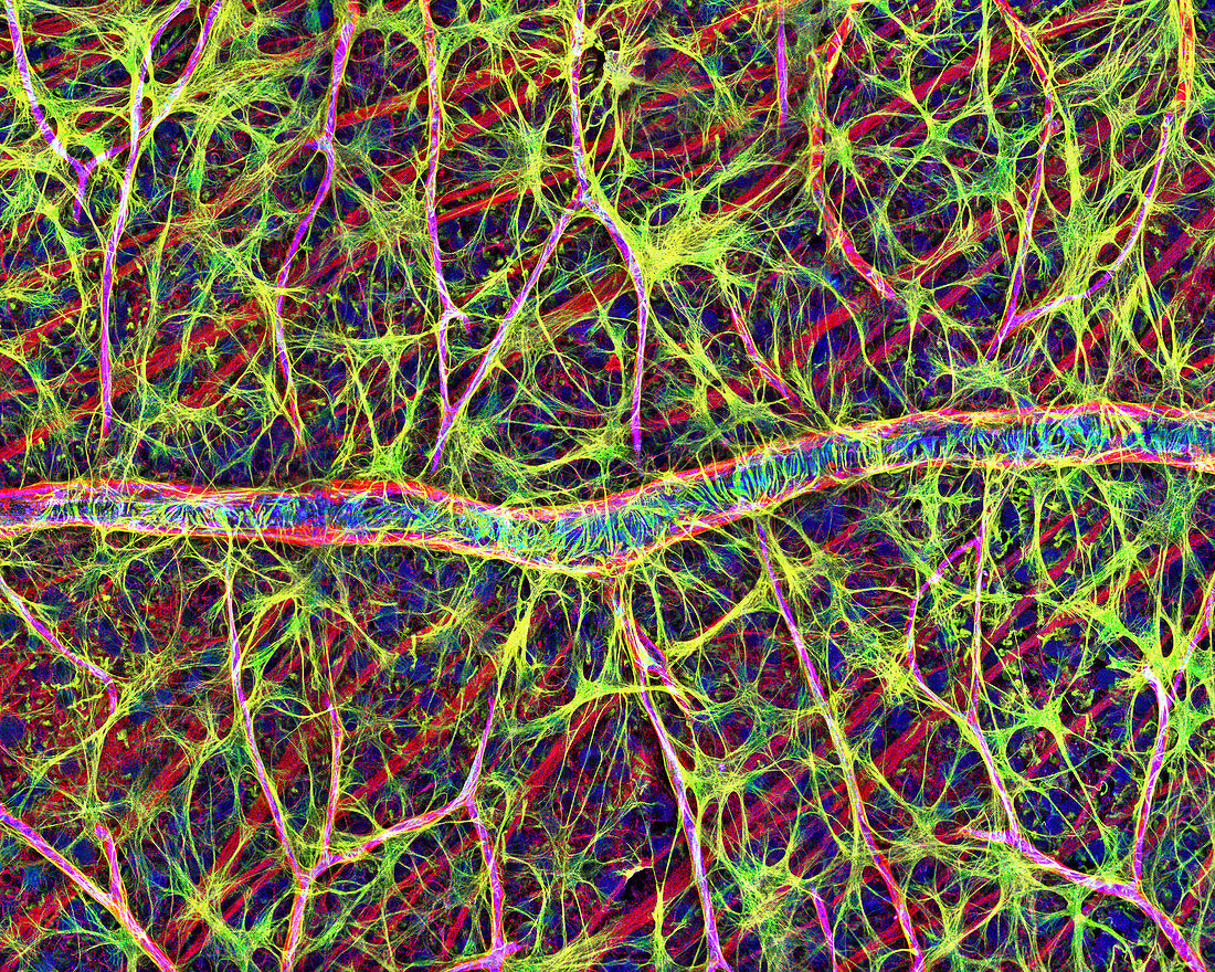
| Retina cells. Fluorescent light micrograph of cells in the retina,the light-sensitive membrane that lines the back of the eyeball. A blood vessel runs from left to right,and numerous other branches are seen. Astrocyte glial cells are yellow. The glial cells provide structural support to the nerve cells that send visual signals to the brain. The tissue has been tagged with fluorescent markers specific to certain proteins. The yellow marks the glial fibrillary acidic protein (GFAP) found in glial cells. Blue marks platelet-endothelial cell adhesion molecule-1 (PECAM-1) found in blood vessels,and red is the structural protein actin. Magnification: x260 when printed 10 cm wide | |
| Lizenzart: | Lizenzpflichtig |
| Credit: | Science Photo Library / Deerinck, Thomas / NCMIR |
| Bildgröße: | 4800 px × 3838 px |
| Modell-Rechte: | nicht erforderlich |
| Eigentums-Rechte: | nicht erforderlich |
| Restrictions: | - |
Preise für dieses Bild ab 15 €
Universitäten & Organisationen
(Informationsmaterial Digital, Informationsmaterial Print, Lehrmaterial Digital etc.)
ab 15 €
Redaktionell
(Bücher, Bücher: Sach- und Fachliteratur, Digitale Medien (redaktionell) etc.)
ab 30 €
Werbung
(Anzeigen, Aussenwerbung, Digitale Medien, Fernsehwerbung, Karten, Werbemittel, Zeitschriften etc.)
ab 55 €
Handelsprodukte
(bedruckte Textilie, Kalender, Postkarte, Grußkarte, Verpackung etc.)
ab 75 €
Pauschalpreise
Rechtepakete für die unbeschränkte Bildnutzung in Print oder Online
ab 495 €
Keywords
- Aktin,
- Anatomie,
- anatomisch,
- Astrozyten,
- Auge,
- Augenheilkunde,
- Biologie,
- biologisch,
- Blutgefäß,
- Blutversorgung,
- Eiweiß,
- Farbstoff,
- Farbstoffe,
- Fluoreszenzlichtmikroskopische Aufnahme,
- gesund,
- GFAP,
- Histologie,
- histologisch,
- kapillar,
- Lichtmikroskop,
- Nervenzelle,
- Netzwerk,
- Neuroglia,
- Neurologie,
- neurologisch,
- Neuron,
- Neuronen,
- normal,
- Retina,
- Sehsinn,
- Sicht,
- Verzweigung,
- Vision