Light micrograph of a section through the eye wall
Bildnummer 11871928
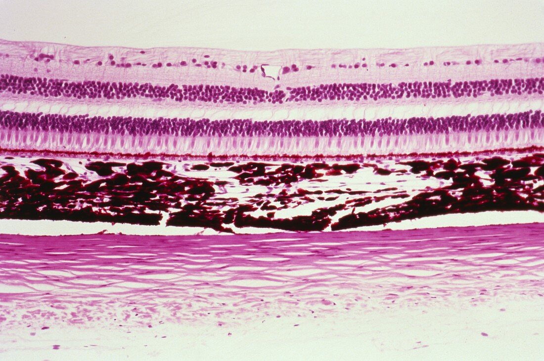
| Eye wall. Light micrograph of a cross section through the eye wall. The outer layer (bottom,pink) is the sclera,connective tissue arranged in bundles. The next layer (centre,dark red) is the choroid. This layer contains blood vessels and a pigment that absorbs excess light to prevent blurred vision. Above the choroid is the layered retina. Its first layer is a line of pigment cells (red),immediately followed by the light sensitive rod and cone cells (pink). For light to reach these cells it must pass through layers of nerve cells. The nerve cell nuclei are visible as the layers of purple dots. Haematoxylin and eosin stained. Magnification x64 at 35mm size | |
| Lizenzart: | Lizenzpflichtig |
| Credit: | Science Photo Library / Biophoto Associates |
| Bildgröße: | 5190 px × 3449 px |
| Modell-Rechte: | nicht erforderlich |
| Eigentums-Rechte: | nicht erforderlich |
| Restrictions: | - |
Preise für dieses Bild ab 15 €
Universitäten & Organisationen
(Informationsmaterial Digital, Informationsmaterial Print, Lehrmaterial Digital etc.)
ab 15 €
Redaktionell
(Bücher, Bücher: Sach- und Fachliteratur, Digitale Medien (redaktionell) etc.)
ab 30 €
Werbung
(Anzeigen, Aussenwerbung, Digitale Medien, Fernsehwerbung, Karten, Werbemittel, Zeitschriften etc.)
ab 55 €
Handelsprodukte
(bedruckte Textilie, Kalender, Postkarte, Grußkarte, Verpackung etc.)
ab 75 €
Pauschalpreise
Rechtepakete für die unbeschränkte Bildnutzung in Print oder Online
ab 495 €