Blood vessels in eye
Bildnummer 11871914
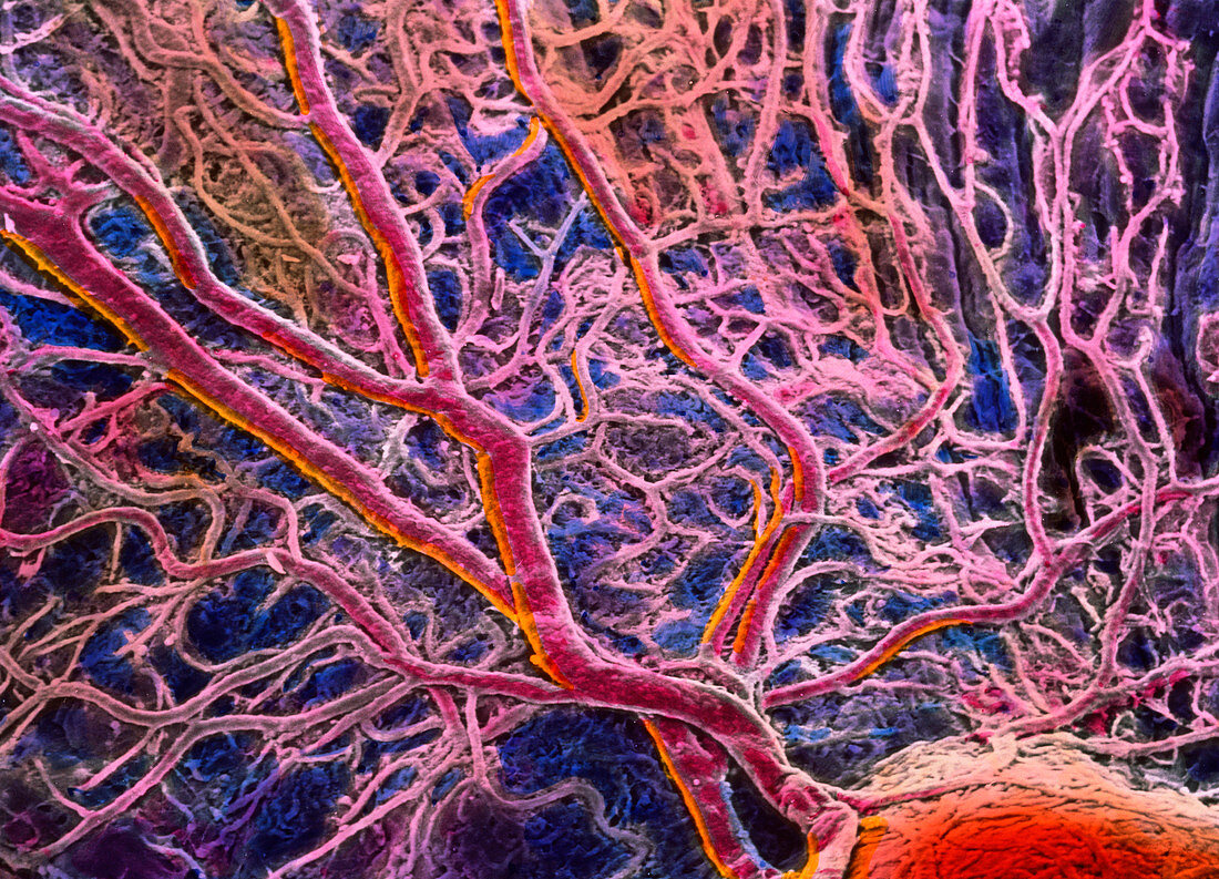
| False-colour scanning electron micrograph (SEM) of blood vessels in the choroid of the eye. A branch- ing network of arteries and veins can be seen in this area under the central fovea. The choroid is a tissue layer beneath the retina which supplies blood,thus food and oxygen,to light sensitive cells of the eye. Many cone cells occur on the retina at the central fovea area,specialized for acute day vision,and hence an area that uses much blood. The choroid is also darkened (blue) with pigment cells; pigment absorbs light rays passing through the retina and prevents light reflection. Magnification: x46 at 6x7cm size. Magnification: x70 at 4x5 inch size | |
| Lizenzart: | Lizenzpflichtig |
| Credit: | Science Photo Library / UNIVERSITY LA SAPIENZA, ROME / DEPT. OF ANATOMY / PROF. P. MOTTA |
| Bildgröße: | 4331 px × 3124 px |
| Modell-Rechte: | nicht erforderlich |
| Eigentums-Rechte: | nicht erforderlich |
| Restrictions: | - |
Preise für dieses Bild ab 15 €
Universitäten & Organisationen
(Informationsmaterial Digital, Informationsmaterial Print, Lehrmaterial Digital etc.)
ab 15 €
Redaktionell
(Bücher, Bücher: Sach- und Fachliteratur, Digitale Medien (redaktionell) etc.)
ab 30 €
Werbung
(Anzeigen, Aussenwerbung, Digitale Medien, Fernsehwerbung, Karten, Werbemittel, Zeitschriften etc.)
ab 55 €
Handelsprodukte
(bedruckte Textilie, Kalender, Postkarte, Grußkarte, Verpackung etc.)
ab 75 €
Pauschalpreise
Rechtepakete für die unbeschränkte Bildnutzung in Print oder Online
ab 495 €