False-colour SEM of the structure of the retina
Bildnummer 11871907
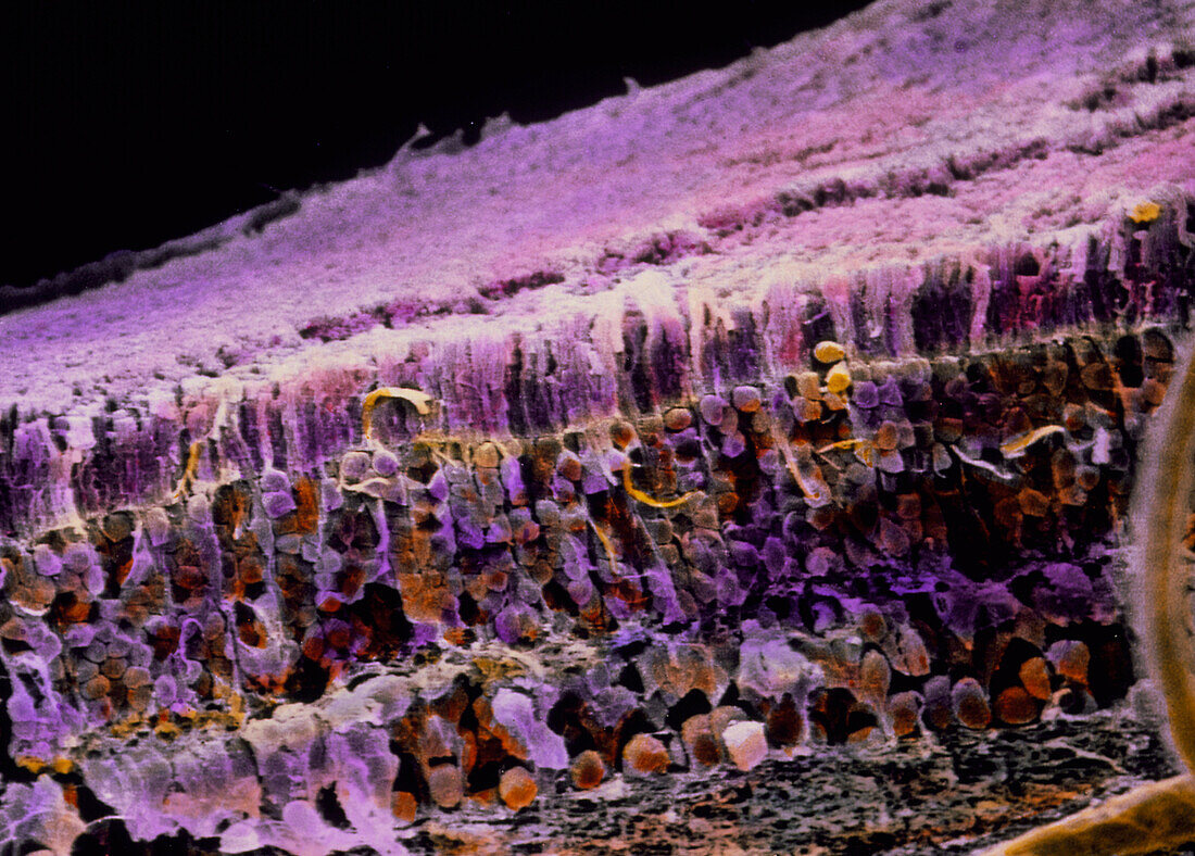
| Retina. Coloured scanning electron micrograph (SEM) of the structure of the eye retina. The light-sensitive retina detects visible images. It consists of many layers of cells,seen here. Light strikes from below. At top is the surface of pigment cells (white). Directly beneath lie close- packed rod and cone cells (pink). More numerous rod cells appear as lighter fibres; cone cells are darker and cone-like. While rod cells detect light intensity,cone cells respond to colour. A deeper layer of round nerve cells transmits impulses from rods and cones to optic nerve fibres (grey,at bottom centre). Magnification: x400 at 6x7cm size. Magnification: x650 at 4x5 inch size | |
| Lizenzart: | Lizenzpflichtig |
| Credit: | Science Photo Library / UNIVERSITY LA SAPIENZA, ROME / DEPT. OF ANATOMY / PROF. P. MOTTA |
| Bildgröße: | 3543 px × 2537 px |
| Modell-Rechte: | nicht erforderlich |
| Eigentums-Rechte: | nicht erforderlich |
| Restrictions: | - |
Preise für dieses Bild ab 15 €
Universitäten & Organisationen
(Informationsmaterial Digital, Informationsmaterial Print, Lehrmaterial Digital etc.)
ab 15 €
Redaktionell
(Bücher, Bücher: Sach- und Fachliteratur, Digitale Medien (redaktionell) etc.)
ab 30 €
Werbung
(Anzeigen, Aussenwerbung, Digitale Medien, Fernsehwerbung, Karten, Werbemittel, Zeitschriften etc.)
ab 55 €
Handelsprodukte
(bedruckte Textilie, Kalender, Postkarte, Grußkarte, Verpackung etc.)
ab 75 €
Pauschalpreise
Rechtepakete für die unbeschränkte Bildnutzung in Print oder Online
ab 495 €