Artwork of eye
Bildnummer 11871900
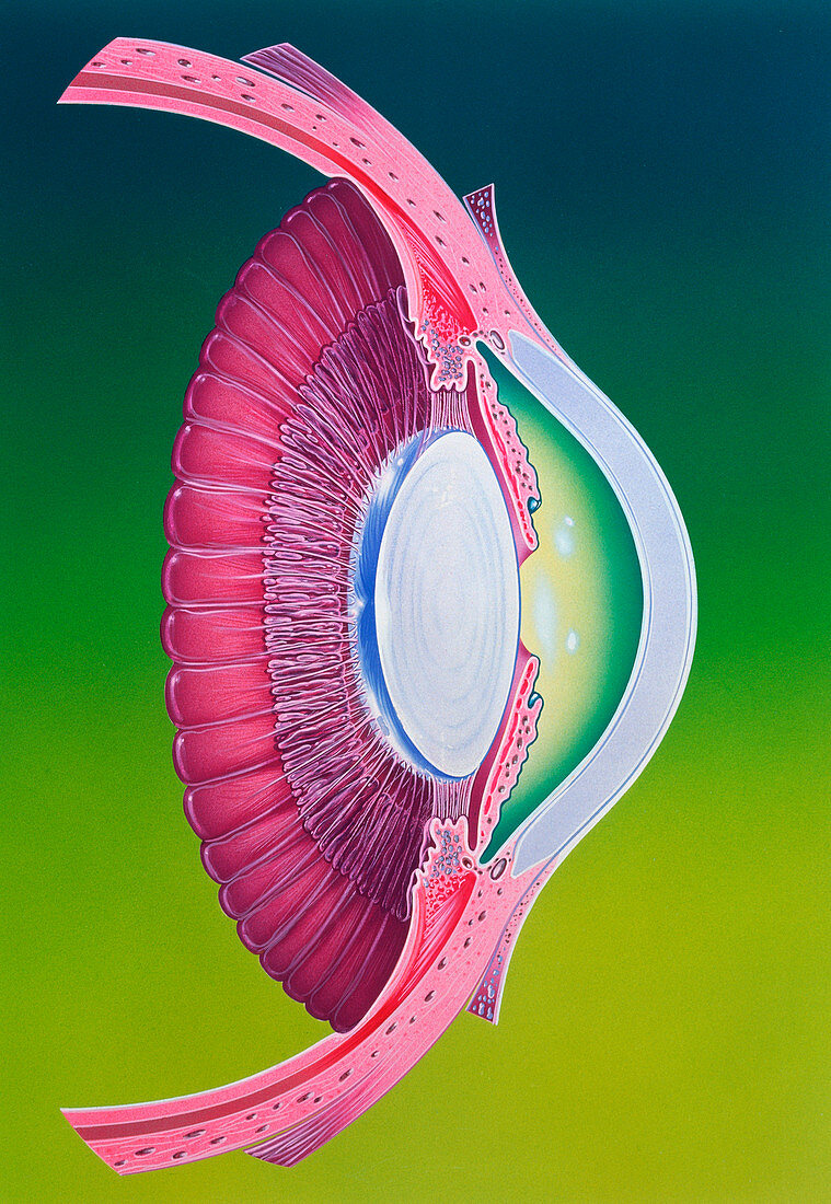
| Illustration of the anterior chamber of the eye. Anterior structures in the eyeball focus an image onto the retina nerve cells at the back of the eye. The cornea (centre right,white) protrudes as the front circular part which serves as the main lens. It performs most of the focusing,and is covered by a thin protective conjunctiva mem- brane. Behind the cornea is a chamber of aqueous humour (fluid) and behind that,a central hole or pupil surrounded by a coloured iris (here purple). Tiny muscles alter the pupil size to control the amount of light entering the eye. In contact with,and behind the iris,is a crystalline lens (at centre,white) providing more focusing power | |
| Lizenzart: | Lizenzpflichtig |
| Credit: | Science Photo Library / Bavosi, John |
| Bildgröße: | 3082 px × 4464 px |
| Modell-Rechte: | nicht erforderlich |
| Eigentums-Rechte: | nicht erforderlich |
| Restrictions: | - |
Preise für dieses Bild ab 15 €
Universitäten & Organisationen
(Informationsmaterial Digital, Informationsmaterial Print, Lehrmaterial Digital etc.)
ab 15 €
Redaktionell
(Bücher, Bücher: Sach- und Fachliteratur, Digitale Medien (redaktionell) etc.)
ab 30 €
Werbung
(Anzeigen, Aussenwerbung, Digitale Medien, Fernsehwerbung, Karten, Werbemittel, Zeitschriften etc.)
ab 55 €
Handelsprodukte
(bedruckte Textilie, Kalender, Postkarte, Grußkarte, Verpackung etc.)
ab 75 €
Pauschalpreise
Rechtepakete für die unbeschränkte Bildnutzung in Print oder Online
ab 495 €