Fundus camera image of a normal retina
Bildnummer 11871886
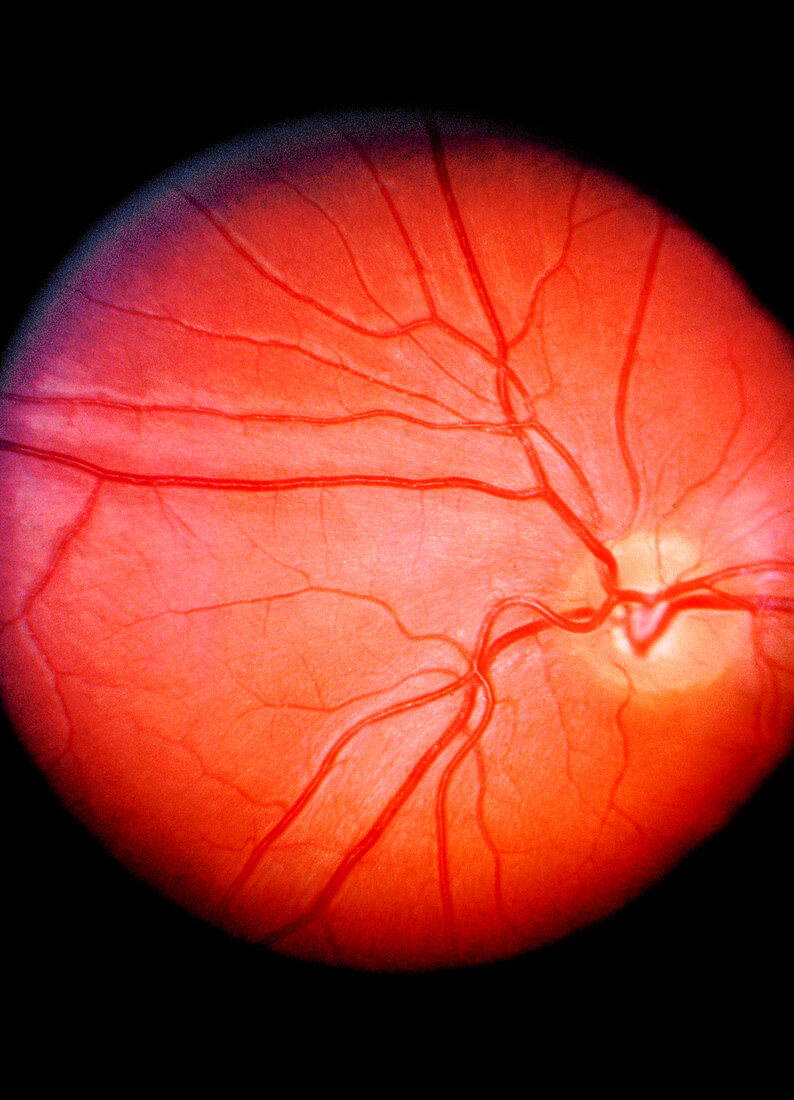
| Fundus camera image of the retina of a normal eye,showing the distribution of the retinal veins & arteries: the central retinal artery (a branch of the opthalmic artery) enters the optic nerve 2 cm before it reaches the eyeball,emerging through the retina in the centre of the optic disc (the blind spot of the eye,the pale area on right). An extension of the optic nerve,the retina consists of photosensitive cells (rods & cones) which translate light energy into nervous impulses. The orange-red appearance is caused by the pigment & abundance of blood vessels in the choroid layer,visible through the normally transparent retina | |
| Lizenzart: | Lizenzpflichtig |
| Credit: | Science Photo Library / John Radcliffe Infirmary |
| Bildgröße: | 3551 px × 4920 px |
| Modell-Rechte: | nicht erforderlich |
| Eigentums-Rechte: | nicht erforderlich |
| Restrictions: | - |
Preise für dieses Bild ab 15 €
Universitäten & Organisationen
(Informationsmaterial Digital, Informationsmaterial Print, Lehrmaterial Digital etc.)
ab 15 €
Redaktionell
(Bücher, Bücher: Sach- und Fachliteratur, Digitale Medien (redaktionell) etc.)
ab 30 €
Werbung
(Anzeigen, Aussenwerbung, Digitale Medien, Fernsehwerbung, Karten, Werbemittel, Zeitschriften etc.)
ab 55 €
Handelsprodukte
(bedruckte Textilie, Kalender, Postkarte, Grußkarte, Verpackung etc.)
ab 75 €
Pauschalpreise
Rechtepakete für die unbeschränkte Bildnutzung in Print oder Online
ab 495 €