Nerve cell growth
Bildnummer 11871294
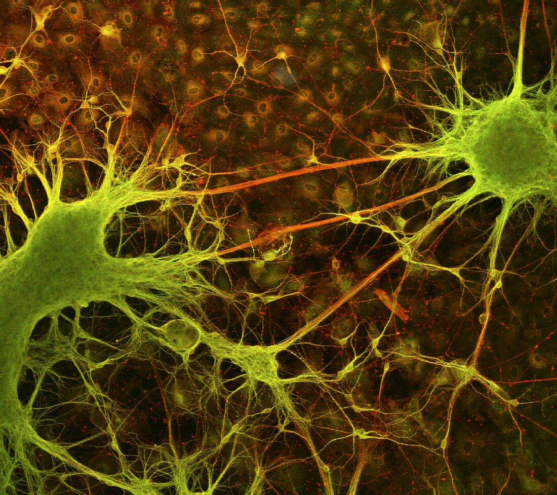
| Nerve cell growth. Light micrograph of nerve cells (neurons) with immunofluorescent staining. These cells have been grown in culture. The stains show neurites (thin strands,either axons or dendrites) connecting nerve cell bodies. Short neurites are green,long ones are red. Two large clusters of nerve cell bodies are at left and right,with non- neuronal cell nuclei in the background. Neurons transmit electrical signals around the body,especially the brain and the spinal cord. These neurons have been grown from NT2 cells,a human teratocarcinoma cell line that can differentiate into nerve cells. Such research may make neural regeneration treatments possible | |
| Lizenzart: | Lizenzpflichtig |
| Credit: | Science Photo Library / Paquet-Durand, Francois |
| Bildgröße: | 2700 px × 2399 px |
| Modell-Rechte: | nicht erforderlich |
| Eigentums-Rechte: | nicht erforderlich |
| Restrictions: | - |
Preise für dieses Bild ab 15 €
Universitäten & Organisationen
(Informationsmaterial Digital, Informationsmaterial Print, Lehrmaterial Digital etc.)
ab 15 €
Redaktionell
(Bücher, Bücher: Sach- und Fachliteratur, Digitale Medien (redaktionell) etc.)
ab 30 €
Werbung
(Anzeigen, Aussenwerbung, Digitale Medien, Fernsehwerbung, Karten, Werbemittel, Zeitschriften etc.)
ab 55 €
Handelsprodukte
(bedruckte Textilie, Kalender, Postkarte, Grußkarte, Verpackung etc.)
ab 75 €
Pauschalpreise
Rechtepakete für die unbeschränkte Bildnutzung in Print oder Online
ab 495 €