Light micrograph of human spinal cord
Bildnummer 11870951
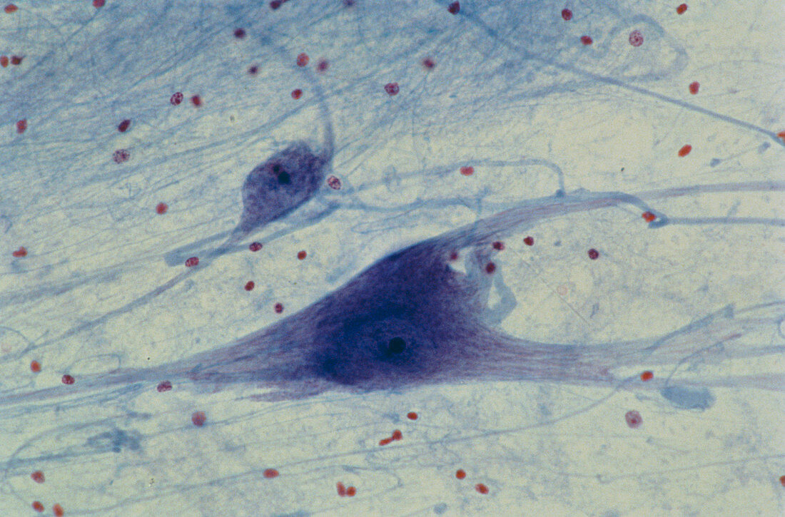
| Light micrograph of normal human spinal cord,showing part of the grey matter of the anterior horn. Two neurons (nerve cells) are visible,each with a dark-stained nucleous and associated with a mass of nerve fibres. The scattered rose-purple particles in the background are the nuclei of neurological cells; their cytoplasm is unstained. They lie within a field of intercrossing nerve fibres,seen as delicate pale blue lines. This region of neurological cells and nerve fibres is known as the neuropil. Magnification x100 at 35mm size | |
| Lizenzart: | Lizenzpflichtig |
| Credit: | Science Photo Library / Michler, Astrid & Hans-Frieder |
| Bildgröße: | 5575 px × 3672 px |
| Modell-Rechte: | nicht erforderlich |
| Eigentums-Rechte: | nicht erforderlich |
| Restrictions: | - |
Preise für dieses Bild ab 15 €
Universitäten & Organisationen
(Informationsmaterial Digital, Informationsmaterial Print, Lehrmaterial Digital etc.)
ab 15 €
Redaktionell
(Bücher, Bücher: Sach- und Fachliteratur, Digitale Medien (redaktionell) etc.)
ab 30 €
Werbung
(Anzeigen, Aussenwerbung, Digitale Medien, Fernsehwerbung, Karten, Werbemittel, Zeitschriften etc.)
ab 55 €
Handelsprodukte
(bedruckte Textilie, Kalender, Postkarte, Grußkarte, Verpackung etc.)
ab 75 €
Pauschalpreise
Rechtepakete für die unbeschränkte Bildnutzung in Print oder Online
ab 495 €