Light micrograph of human spinal cord
Bildnummer 11870950
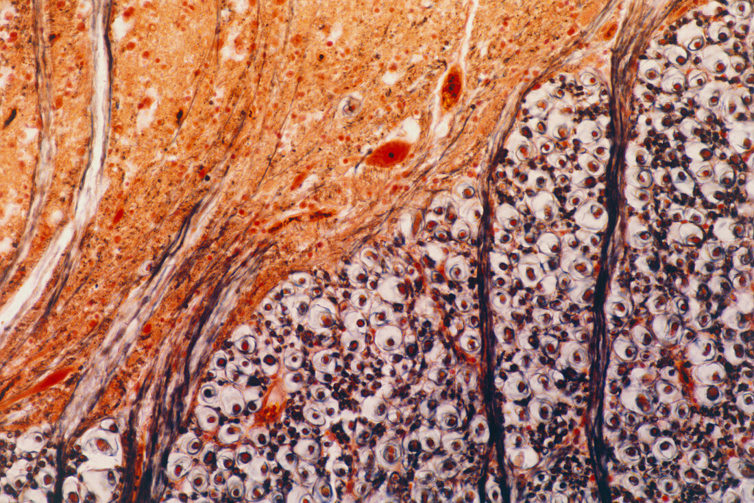
| Light micrograph of normal human spinal cord,showing the junction between the grey matter (bottom right) and the white matter (top left) in the anterior horn. The large red cells (two visible) are anterior horn cells,embedded in a mass of neuroglial cells,nerve fibres (purple lines) and capillaries (unstained vessel,bottom right). The bulk of the white matter consists of cross sectioned myelinated nerve fibres seen as a clear ring (myelin) surrounding a central dot (the fibre). Fibre bundles from the grey matter (vertical lines,left) are seen in longitudinal section. Magnification X 50 at 35mm size | |
| Lizenzart: | Lizenzpflichtig |
| Credit: | Science Photo Library / Michler, Astrid & Hans-Frieder |
| Bildgröße: | 5433 px × 3631 px |
| Modell-Rechte: | nicht erforderlich |
| Eigentums-Rechte: | nicht erforderlich |
| Restrictions: | - |
Preise für dieses Bild ab 15 €
Universitäten & Organisationen
(Informationsmaterial Digital, Informationsmaterial Print, Lehrmaterial Digital etc.)
ab 15 €
Redaktionell
(Bücher, Bücher: Sach- und Fachliteratur, Digitale Medien (redaktionell) etc.)
ab 30 €
Werbung
(Anzeigen, Aussenwerbung, Digitale Medien, Fernsehwerbung, Karten, Werbemittel, Zeitschriften etc.)
ab 55 €
Handelsprodukte
(bedruckte Textilie, Kalender, Postkarte, Grußkarte, Verpackung etc.)
ab 75 €
Pauschalpreise
Rechtepakete für die unbeschränkte Bildnutzung in Print oder Online
ab 495 €