Light micrograph of human spinal cord
Bildnummer 11870949
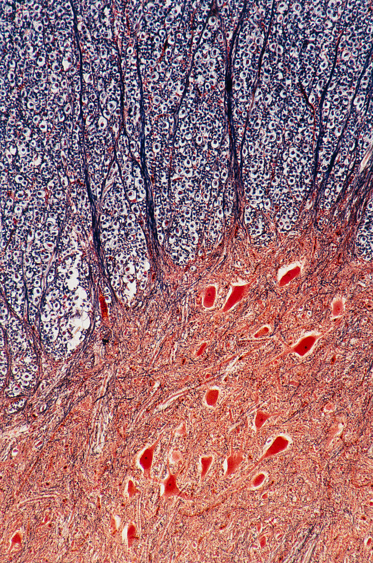
| Light micrograph of normal human spinal cord. This cross section shows the junction between the grey matter (bottom,orange) and the white matter (top) in the anterior horn. The large cells,red,(some with pale nucleus and dense nucleolus) are anterior horn cells,whose axons become the efferent root fibres. They are embedded in a mass of neuroglial cells. In cross section (most of upper field) they appear as a clear ring (myelin) around a central dot (the fibres). The prominent ascending lines are fibres cut longitudinally. Magnification: X 20 at 35mm size | |
| Lizenzart: | Lizenzpflichtig |
| Credit: | Science Photo Library / Michler, Astrid & Hans-Frieder |
| Bildgröße: | 3535 px × 5326 px |
| Modell-Rechte: | nicht erforderlich |
| Eigentums-Rechte: | nicht erforderlich |
| Restrictions: | - |
Preise für dieses Bild ab 15 €
Universitäten & Organisationen
(Informationsmaterial Digital, Informationsmaterial Print, Lehrmaterial Digital etc.)
ab 15 €
Redaktionell
(Bücher, Bücher: Sach- und Fachliteratur, Digitale Medien (redaktionell) etc.)
ab 30 €
Werbung
(Anzeigen, Aussenwerbung, Digitale Medien, Fernsehwerbung, Karten, Werbemittel, Zeitschriften etc.)
ab 55 €
Handelsprodukte
(bedruckte Textilie, Kalender, Postkarte, Grußkarte, Verpackung etc.)
ab 75 €
Pauschalpreise
Rechtepakete für die unbeschränkte Bildnutzung in Print oder Online
ab 495 €