Image-processing brain activity
Bildnummer 11870933
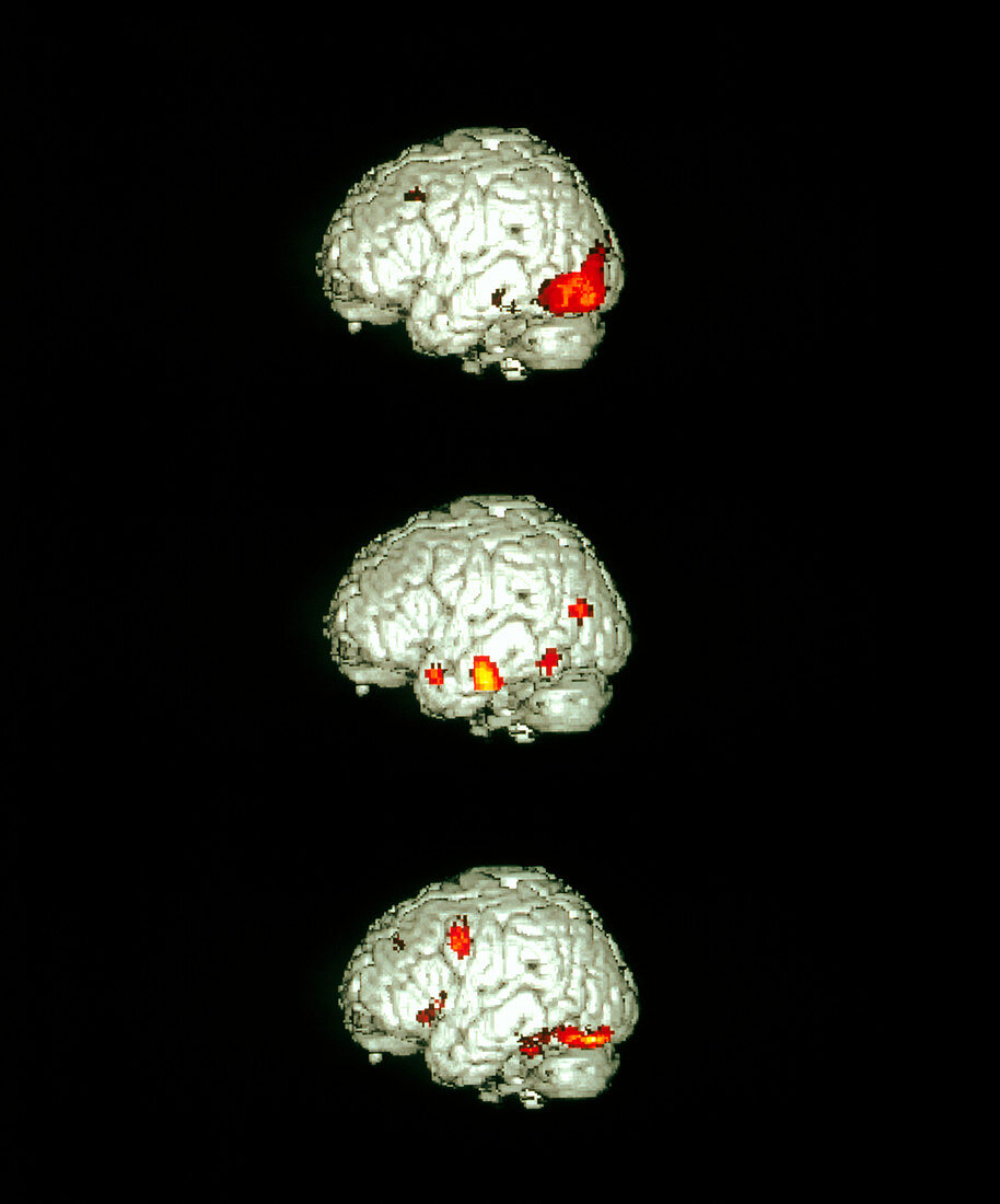
| Image-processing brain activity. Coloured positron emission tomography (PET) scans of the human brain areas active while processing images of objects. The left sides of the brains are seen,with active parts red/orange. At top,the visual area of the occipital cortex is activated as objects are seen. At centre,areas within the temporal lobe light up as objects are recognised. At bottom,speech and motor areas within the frontal cortex (centre left of scan) are activated as objects are named. The scans involved injecting radioactive oxygen-15 into the bloodstream so that the areas of the brain with high metabolic activity can be seen | |
| Lizenzart: | Lizenzpflichtig |
| Credit: | Science Photo Library / Wellcome Dept. of Cognitive Neurology |
| Bildgröße: | 2943 px × 3543 px |
| Modell-Rechte: | nicht erforderlich |
| Eigentums-Rechte: | nicht erforderlich |
| Restrictions: | - |
Preise für dieses Bild ab 15 €
Universitäten & Organisationen
(Informationsmaterial Digital, Informationsmaterial Print, Lehrmaterial Digital etc.)
ab 15 €
Redaktionell
(Bücher, Bücher: Sach- und Fachliteratur, Digitale Medien (redaktionell) etc.)
ab 30 €
Werbung
(Anzeigen, Aussenwerbung, Digitale Medien, Fernsehwerbung, Karten, Werbemittel, Zeitschriften etc.)
ab 55 €
Handelsprodukte
(bedruckte Textilie, Kalender, Postkarte, Grußkarte, Verpackung etc.)
ab 75 €
Pauschalpreise
Rechtepakete für die unbeschränkte Bildnutzung in Print oder Online
ab 495 €