Functional map of the brain's visual cortex areas
Bildnummer 11870922
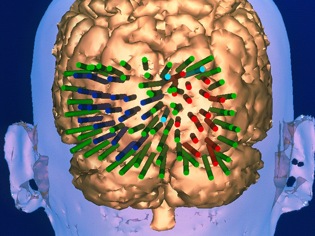
| Visual cortex brain areas. 3-D magnetic resonance imaging (MRI) scan of the brain mapped to reveal areas of the cerebral visual cortex,which controls vision. This mapping is used in virtual reality assisted surgery. Electric currents are first applied to parts of the visual cortex to supress vision and create a map. The map is shown by colour-coded cylinders: red (vision suppressed to right visual field),blue (vision suppressed to left visual field) and green (no effect). By knowing these regions,surgery such as tumour removal can be done with minimum damage to the visual cortex | |
| Lizenzart: | Lizenzpflichtig |
| Credit: | Science Photo Library / BRIGHAM & WOMEN'S HOSPITAL / SURGICAL PLANNING LAB / MIT AI LAB |
| Bildgröße: | 3543 px × 2653 px |
| Modell-Rechte: | nicht erforderlich |
| Eigentums-Rechte: | nicht erforderlich |
| Restrictions: | - |
Preise für dieses Bild ab 15 €
Universitäten & Organisationen
(Informationsmaterial Digital, Informationsmaterial Print, Lehrmaterial Digital etc.)
ab 15 €
Redaktionell
(Bücher, Bücher: Sach- und Fachliteratur, Digitale Medien (redaktionell) etc.)
ab 30 €
Werbung
(Anzeigen, Aussenwerbung, Digitale Medien, Fernsehwerbung, Karten, Werbemittel, Zeitschriften etc.)
ab 55 €
Handelsprodukte
(bedruckte Textilie, Kalender, Postkarte, Grußkarte, Verpackung etc.)
ab 75 €
Pauschalpreise
Rechtepakete für die unbeschränkte Bildnutzung in Print oder Online
ab 495 €