PET brain scan amygdala response to fear
Bildnummer 11870919
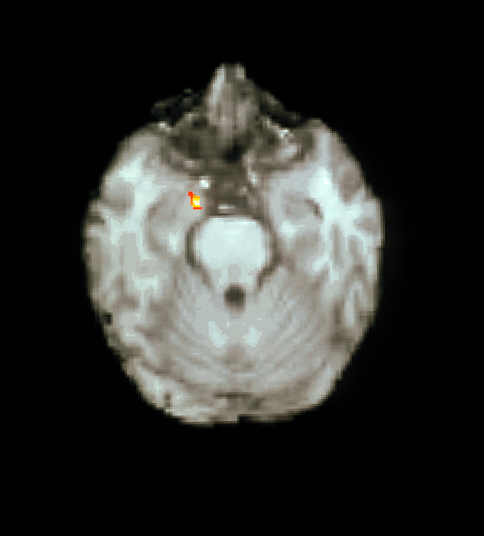
| Brain response to fear. Coloured positron emission tomography (PET) scan of a transverse section through a human brain,showing response to fear. The active brain region colour-coded yellow/red is the left amygdala (at upper centre left). Subjects in this study viewed a series of photographs of a man's face depicting increasing stages of fearful expression (see photo M245/349). With increased fearful expression the amygdala response increases suggesting it is this part of the brain that recognises and responds to facial fear. PET scanning detects blood flow & metabolic activity; here the background brain is an MRI image | |
| Lizenzart: | Lizenzpflichtig |
| Credit: | Science Photo Library / Wellcome Dept. of Cognitive Neurology |
| Bildgröße: | 3201 px × 3543 px |
| Modell-Rechte: | nicht erforderlich |
| Eigentums-Rechte: | nicht erforderlich |
| Restrictions: | - |
Preise für dieses Bild ab 15 €
Universitäten & Organisationen
(Informationsmaterial Digital, Informationsmaterial Print, Lehrmaterial Digital etc.)
ab 15 €
Redaktionell
(Bücher, Bücher: Sach- und Fachliteratur, Digitale Medien (redaktionell) etc.)
ab 30 €
Werbung
(Anzeigen, Aussenwerbung, Digitale Medien, Fernsehwerbung, Karten, Werbemittel, Zeitschriften etc.)
ab 55 €
Handelsprodukte
(bedruckte Textilie, Kalender, Postkarte, Grußkarte, Verpackung etc.)
ab 75 €
Pauschalpreise
Rechtepakete für die unbeschränkte Bildnutzung in Print oder Online
ab 495 €