Ventricles of brain,MRI
Bildnummer 11870808
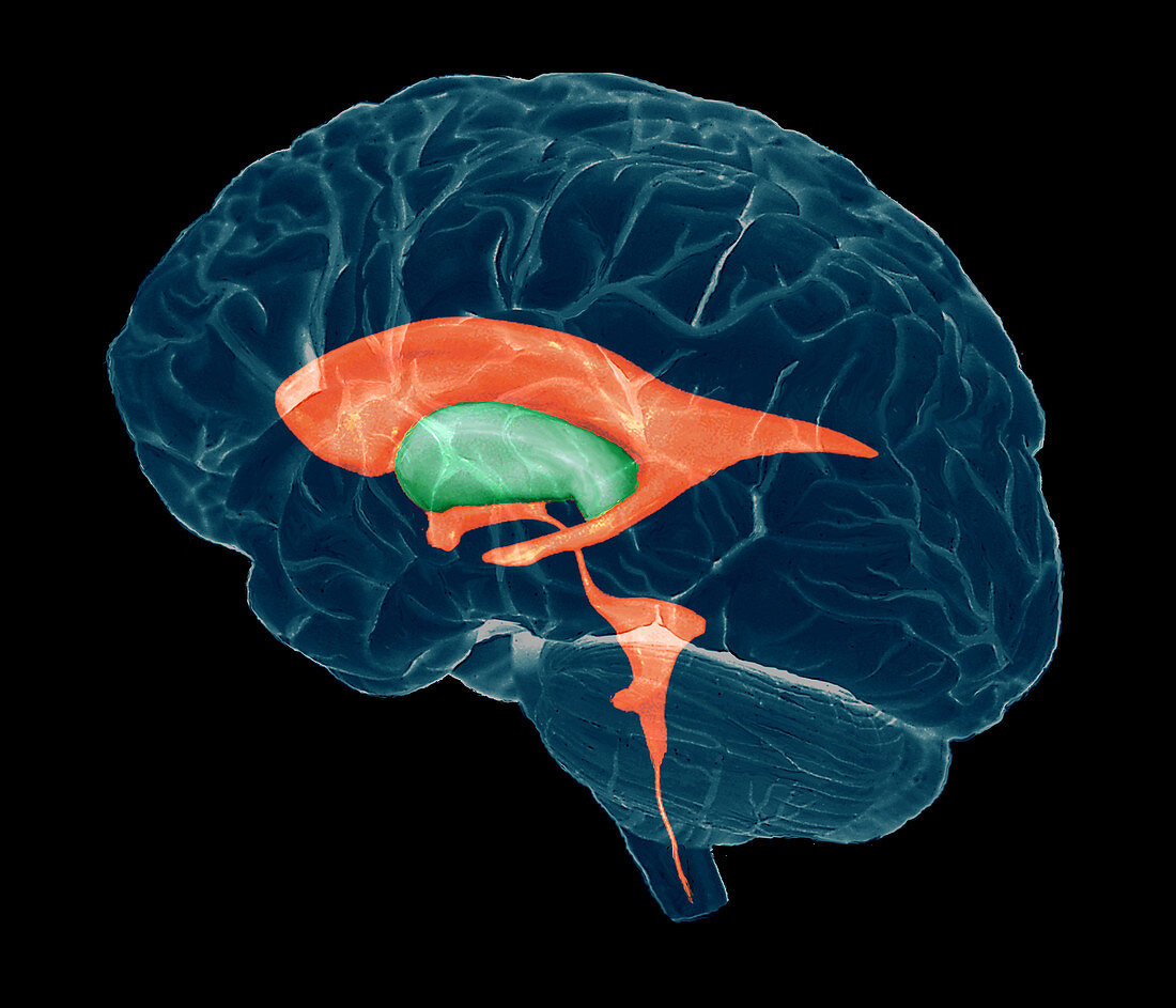
| Ventricles of brain. Coloured composite 3-D magnetic resonance imaging (MRI) scan of a brain in profile,showing the ventricles (red). The front of the brain is at left. Cushioning cerebrospinal fluid (CSF) circulates in the ventricles and runs down the brainstem (bottom right) to the spinal cord. The curved lateral ventricles (one seen) lie on either side of the brain,one in each cerebral hemisphere. They communicate with the small third ventricle (lower centre),which lies between and below them. This leads down to the fourth ventricle (lower right) via a channel,the cerebral aqueduct. One of the sensory-processing thalami (green) is also seen | |
| Lizenzart: | Lizenzpflichtig |
| Credit: | Science Photo Library / Zephyr |
| Bildgröße: | 4134 px × 3543 px |
| Modell-Rechte: | nicht erforderlich |
| Eigentums-Rechte: | nicht erforderlich |
| Restrictions: | - |
Preise für dieses Bild ab 15 €
Universitäten & Organisationen
(Informationsmaterial Digital, Informationsmaterial Print, Lehrmaterial Digital etc.)
ab 15 €
Redaktionell
(Bücher, Bücher: Sach- und Fachliteratur, Digitale Medien (redaktionell) etc.)
ab 30 €
Werbung
(Anzeigen, Aussenwerbung, Digitale Medien, Fernsehwerbung, Karten, Werbemittel, Zeitschriften etc.)
ab 55 €
Handelsprodukte
(bedruckte Textilie, Kalender, Postkarte, Grußkarte, Verpackung etc.)
ab 75 €
Pauschalpreise
Rechtepakete für die unbeschränkte Bildnutzung in Print oder Online
ab 495 €
Keywords
- 3-d,
- 3D,
- Anatomie,
- Aquädukte,
- Bild,
- csf,
- Dreidimensional,
- Falten,
- farbig,
- Gefaltet,
- Gehirn,
- gesund,
- Hemisphären,
- Hirnstamm,
- Hohlraum,
- Kleinhirn,
- Kreislauf,
- Magnetresonanztomografie,
- menschlicher Körper,
- MRI,
- neural,
- Neuroimaging,
- Neurologie,
- normal,
- Scan,
- Scannen,
- seitlich,
- sensorisch,
- Sinn,
- Sinne,
- System,
- Thalamus,
- Ventrikel,
- wird bearbeitet,
- zentrales Nervensystem,
- Zusammengesetzt