Coloured 3-D MRI scan of brain in CT scan of skull
Bildnummer 11870767
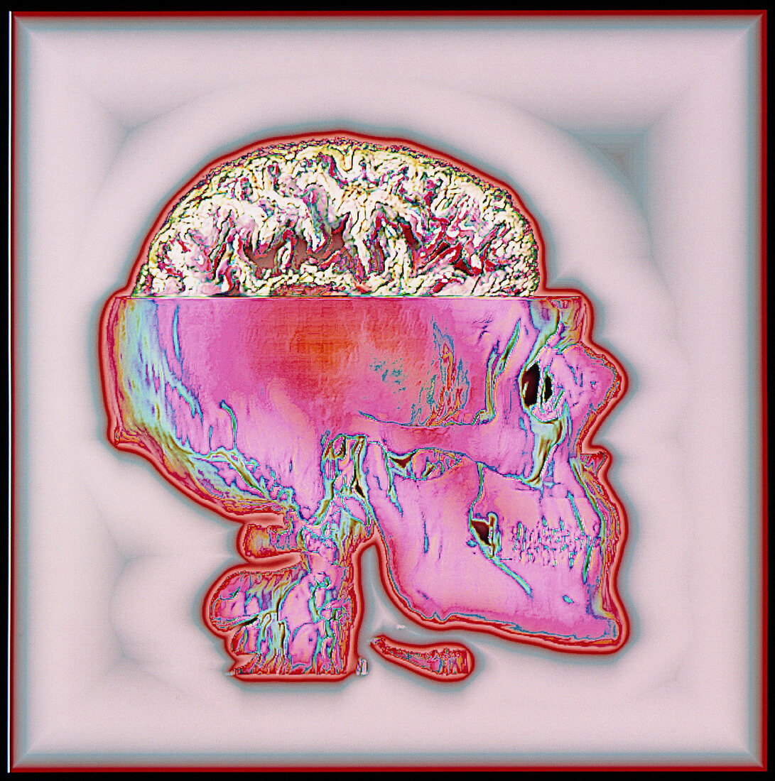
| Brain in skull. Coloured three-dimensional magnetic resonance imaging (MRI) scan of a healthy human brain (white & brown). It is seen in a coloured three-dimensional (3-D) computer tomography (CT) scan of a skull (pink). The front of the skull is at right. The folded cerebrum,the largest part of the brain and centre of conscious brain activity,is seen. MRI scanners use pulses of radio waves to build slice images of the human body. CT scanning also produces slice images,but by using X-rays instead. Several slice images were put together to form these 3-D MRI and CT scans using a computer before they were combined | |
| Lizenzart: | Lizenzpflichtig |
| Credit: | Science Photo Library / Tsiaras, Alexander |
| Bildgröße: | 3777 px × 3807 px |
| Modell-Rechte: | nicht erforderlich |
| Eigentums-Rechte: | nicht erforderlich |
| Restrictions: |
|
Preise für dieses Bild ab 15 €
Universitäten & Organisationen
(Informationsmaterial Digital, Informationsmaterial Print, Lehrmaterial Digital etc.)
ab 15 €
Redaktionell
(Bücher, Bücher: Sach- und Fachliteratur, Digitale Medien (redaktionell) etc.)
ab 30 €
Werbung
(Anzeigen, Aussenwerbung, Digitale Medien, Fernsehwerbung, Karten, Werbemittel, Zeitschriften etc.)
ab 55 €
Handelsprodukte
(bedruckte Textilie, Kalender, Postkarte, Grußkarte, Verpackung etc.)
ab 75 €
Pauschalpreise
Rechtepakete für die unbeschränkte Bildnutzung in Print oder Online
ab 495 €