NMR scan of an infant's head,sagittal section
Bildnummer 11870674
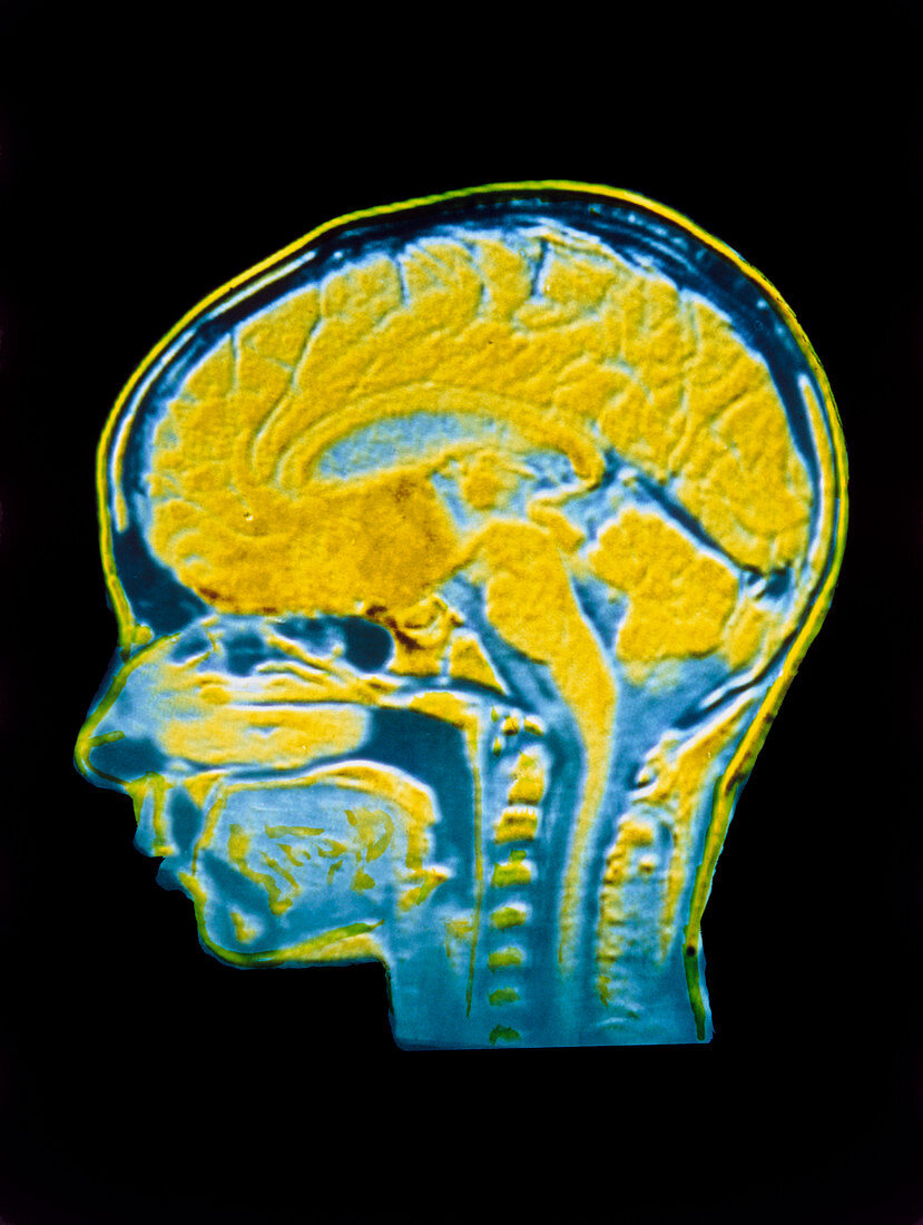
| False-colour nuclear magnetic resonance (NMR) image of a sagittal section through the head of a human infant,showing structures of a normal brain. The cerebrum (the 2 cerebral hemispheres) which forms the bulk of brain tissue appears yellow. The blue areas in centre and surrounding the spinal cord at bottom represent cerebro-spinal fluid (CSF); the central blue area shows CSF in the ventricles (chambers) at the centre of the brain. The spinal cord is clearly visible running down the centre of neck (vertebrae are visible on either side),arising from the brainstem,the swelling at the top of the spinal cord | |
| Lizenzart: | Lizenzpflichtig |
| Credit: | Science Photo Library / CNRI |
| Bildgröße: | 3265 px × 4333 px |
| Modell-Rechte: | nicht erforderlich |
| Eigentums-Rechte: | nicht erforderlich |
| Restrictions: | - |
Preise für dieses Bild ab 15 €
Universitäten & Organisationen
(Informationsmaterial Digital, Informationsmaterial Print, Lehrmaterial Digital etc.)
ab 15 €
Redaktionell
(Bücher, Bücher: Sach- und Fachliteratur, Digitale Medien (redaktionell) etc.)
ab 30 €
Werbung
(Anzeigen, Aussenwerbung, Digitale Medien, Fernsehwerbung, Karten, Werbemittel, Zeitschriften etc.)
ab 55 €
Handelsprodukte
(bedruckte Textilie, Kalender, Postkarte, Grußkarte, Verpackung etc.)
ab 75 €
Pauschalpreise
Rechtepakete für die unbeschränkte Bildnutzung in Print oder Online
ab 495 €