False-colour NMR scan of the head,axial section
Bildnummer 11870670
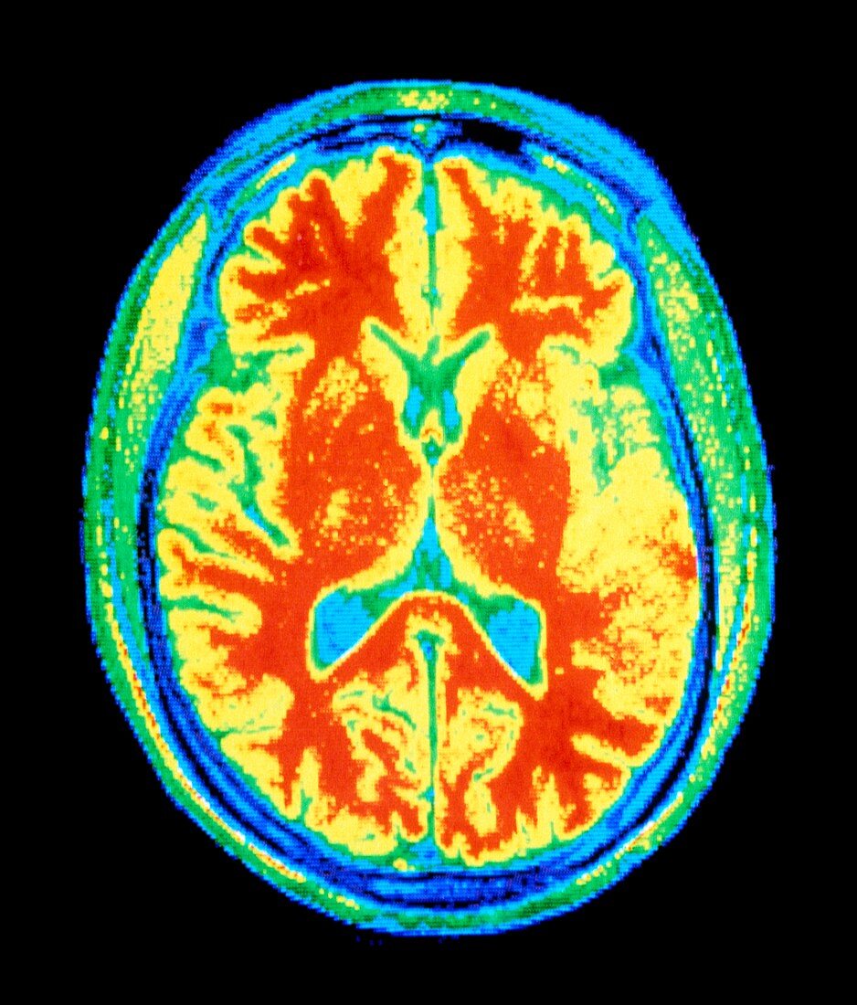
| False-colour nuclear magnetic resonance (NMR) image of an axial section through a human head,showing a normal brain. Fat under the scalp & the underlying bone of the skull appear as various shades of green & yellow. Colour-coding is used to emphasise the division of the brain into outer grey matter (coloured yellow) and inner white matter (coloured red). The level of this section reveals the division of the cerebrum into the left & right cerebral hemispheres. The blue-green areas (centre) represent the fluid-filled ventricles,cavities within the brain | |
| Lizenzart: | Lizenzpflichtig |
| Credit: | Science Photo Library / CNRI |
| Bildgröße: | 3899 px × 4571 px |
| Modell-Rechte: | nicht erforderlich |
| Eigentums-Rechte: | nicht erforderlich |
| Restrictions: | - |
Preise für dieses Bild ab 15 €
Universitäten & Organisationen
(Informationsmaterial Digital, Informationsmaterial Print, Lehrmaterial Digital etc.)
ab 15 €
Redaktionell
(Bücher, Bücher: Sach- und Fachliteratur, Digitale Medien (redaktionell) etc.)
ab 30 €
Werbung
(Anzeigen, Aussenwerbung, Digitale Medien, Fernsehwerbung, Karten, Werbemittel, Zeitschriften etc.)
ab 55 €
Handelsprodukte
(bedruckte Textilie, Kalender, Postkarte, Grußkarte, Verpackung etc.)
ab 75 €
Pauschalpreise
Rechtepakete für die unbeschränkte Bildnutzung in Print oder Online
ab 495 €