NMR scan of a child's head,sagittal section
Bildnummer 11870662
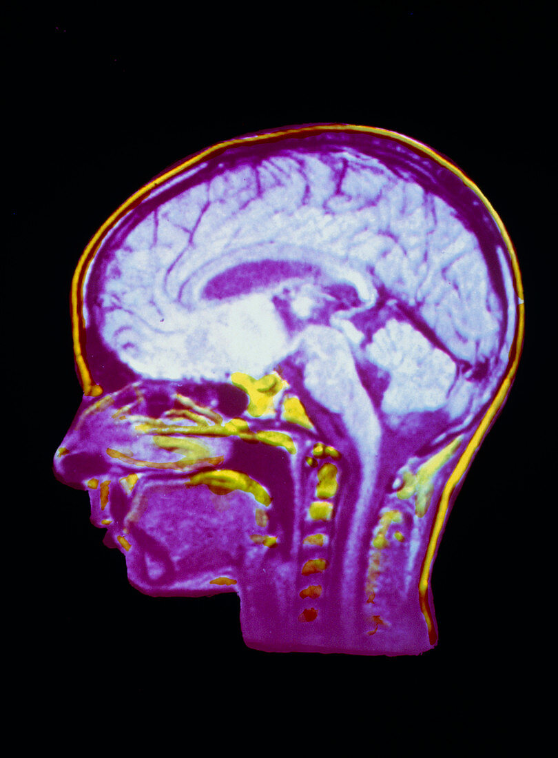
| Nuclear Magnetic Resonance (NMR) image of a sagittal (mid-profile) section through a child's head,showing structures of the brain and spinal cord. Details of the brain visible include the folds of the cerebral cortex,the cerebellum (below the rear lobes of the brain hemispheres) and the pons and medulla of the brainstem,the areas of swelling at the top of the spinal cord,which can be seen running down the neck. Unlike X- ray (CT) scanning,NMR imaging does not use ionising radiations; an image is obtained by studying the radio-response of protons in body tissues that are subjected to a strong,pulsed magnetic field | |
| Lizenzart: | Lizenzpflichtig |
| Credit: | Science Photo Library / CNRI |
| Bildgröße: | 3599 px × 4890 px |
| Modell-Rechte: | nicht erforderlich |
| Eigentums-Rechte: | nicht erforderlich |
| Restrictions: | - |
Preise für dieses Bild ab 15 €
Universitäten & Organisationen
(Informationsmaterial Digital, Informationsmaterial Print, Lehrmaterial Digital etc.)
ab 15 €
Redaktionell
(Bücher, Bücher: Sach- und Fachliteratur, Digitale Medien (redaktionell) etc.)
ab 30 €
Werbung
(Anzeigen, Aussenwerbung, Digitale Medien, Fernsehwerbung, Karten, Werbemittel, Zeitschriften etc.)
ab 55 €
Handelsprodukte
(bedruckte Textilie, Kalender, Postkarte, Grußkarte, Verpackung etc.)
ab 75 €
Pauschalpreise
Rechtepakete für die unbeschränkte Bildnutzung in Print oder Online
ab 495 €