MRI scan of healthy human brain
Bildnummer 11870656
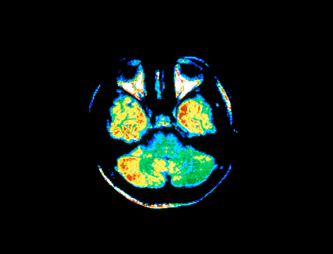
| False colour Nuclear Magnetic Resonance (NMR) image of an adult human brain. The axial section shows parts of lower temporal cerebral lobes (left & right). Above each are the eyes; vitreous humour inside each eyeball appears dark blue. The cerebellum (which consists of 2 cerebellar hemispheres of smoother appearance than the cerebrum) is visible at bottom; it is involved in coordination of muscular activity. The blue & green region immediately above the cerebellum is the pons,a structure of the brainstem (essentially a swelling at the top of the spinal cord),which contains centres for cardiac & respiratory control | |
| Lizenzart: | Lizenzpflichtig |
| Credit: | Science Photo Library / CNRI |
| Bildgröße: | 5088 px × 3874 px |
| Modell-Rechte: | nicht erforderlich |
| Eigentums-Rechte: | nicht erforderlich |
| Restrictions: | - |
Preise für dieses Bild ab 15 €
Universitäten & Organisationen
(Informationsmaterial Digital, Informationsmaterial Print, Lehrmaterial Digital etc.)
ab 15 €
Redaktionell
(Bücher, Bücher: Sach- und Fachliteratur, Digitale Medien (redaktionell) etc.)
ab 30 €
Werbung
(Anzeigen, Aussenwerbung, Digitale Medien, Fernsehwerbung, Karten, Werbemittel, Zeitschriften etc.)
ab 55 €
Handelsprodukte
(bedruckte Textilie, Kalender, Postkarte, Grußkarte, Verpackung etc.)
ab 75 €
Pauschalpreise
Rechtepakete für die unbeschränkte Bildnutzung in Print oder Online
ab 495 €