Light micrograph of a normal thymus
Bildnummer 11869888
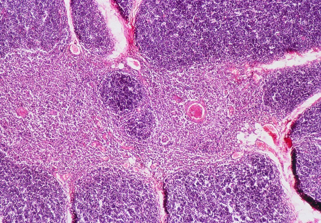
| Light micrograph of normal human thymus. The thymus is divided into lobules separated by septa of connective tissue (white spaces) which may contain blood vessels (as at right,stained red and very dark purple). Each lobule has a cortex (granular masses,densley packed,picture periphery) of lymphocytes,and a medulla (centre) of lymphocytes and stellate epitherial cells. The circular pink structures within the medulla (one prominent) are concentrically arranged epitherial cells of uncertain function. Magnificaton: x20 at 35mm size | |
| Lizenzart: | Lizenzpflichtig |
| Credit: | Science Photo Library / Michler, Astrid & Hans-Frieder |
| Bildgröße: | 5079 px × 3530 px |
| Modell-Rechte: | nicht erforderlich |
| Eigentums-Rechte: | nicht erforderlich |
| Restrictions: | - |
Preise für dieses Bild ab 15 €
Universitäten & Organisationen
(Informationsmaterial Digital, Informationsmaterial Print, Lehrmaterial Digital etc.)
ab 15 €
Redaktionell
(Bücher, Bücher: Sach- und Fachliteratur, Digitale Medien (redaktionell) etc.)
ab 30 €
Werbung
(Anzeigen, Aussenwerbung, Digitale Medien, Fernsehwerbung, Karten, Werbemittel, Zeitschriften etc.)
ab 55 €
Handelsprodukte
(bedruckte Textilie, Kalender, Postkarte, Grußkarte, Verpackung etc.)
ab 75 €
Pauschalpreise
Rechtepakete für die unbeschränkte Bildnutzung in Print oder Online
ab 495 €