Heart,3D CT scans
Bildnummer 11869189
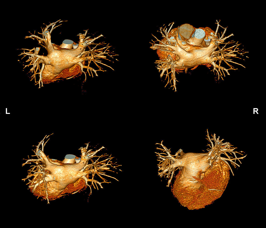
| Heart. Series of coloured 3D CT (computed tomography) scans of the normal heart of a 46-year-old patient. These images show different views of the heart and its blood vessels. The left and right pulmonary arteries branch from the top of the heart from where they carry deoxygenated blood to the lungs. The arteries branch into progressively smaller vessels. The smallest vessels wrap around tiny air sacs (alveoli),which are the site of gaseous exchange. Oxygen enters the blood and carbon dioxide leaves. The oxygenated blood then travels through the pulmonary veins (not seen) back to the heart where it is pumped around the body. This heart is now healthy but was previously treated for arrhythmia (irregular heartbeats) | |
| Lizenzart: | Lizenzpflichtig |
| Credit: | Science Photo Library / Zephyr |
| Bildgröße: | 3969 px × 3402 px |
| Modell-Rechte: | nicht erforderlich |
| Eigentums-Rechte: | nicht erforderlich |
| Restrictions: | - |
Preise für dieses Bild ab 15 €
Universitäten & Organisationen
(Informationsmaterial Digital, Informationsmaterial Print, Lehrmaterial Digital etc.)
ab 15 €
Redaktionell
(Bücher, Bücher: Sach- und Fachliteratur, Digitale Medien (redaktionell) etc.)
ab 30 €
Werbung
(Anzeigen, Aussenwerbung, Digitale Medien, Fernsehwerbung, Karten, Werbemittel, Zeitschriften etc.)
ab 55 €
Handelsprodukte
(bedruckte Textilie, Kalender, Postkarte, Grußkarte, Verpackung etc.)
ab 75 €
Pauschalpreise
Rechtepakete für die unbeschränkte Bildnutzung in Print oder Online
ab 495 €
Keywords
- 3 dimensional,
- 3-d,
- 3-dimensional,
- 3D,
- 40er Jahre,
- Anatomie,
- anatomisch,
- behandelt,
- Biologie,
- biologisch,
- Blutgefäße,
- Computertomographie,
- CT-Scan,
- CT-Scanner,
- Dreidimensional,
- Erwachsene,
- farbig,
- geduldig,
- gefärbt,
- gesund,
- Herz,
- Kardiologie,
- Medizin,
- medizinisch,
- Mensch,
- Menschen,
- menschlicher Körper,
- Muskel,
- normal,
- Organ,
- Person,
- pulmonal,
- Quartett,
- Radiographie,
- repariert,
- Röntgen,
- Röntgengerät,
- Scanner,
- Thorax,
- Truhe,
- vier,
- Vierziger Jahre