Artery tissue layers
Bildnummer 11868907
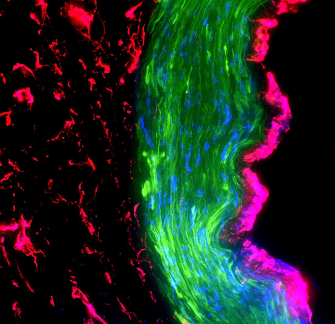
| Artery tissue layers,fluorescence deconvolution micrograph. Fluorescent dyes have been used to highlight tissues and cellular structures: microvasculature (red),smooth muscle (green),cell nuclei (blue),intima (pink). The layers seen here are the tunica intima (right,pink,the internal layer),the tunica media (centre,green,the middle layer) and the tunica adventitia (left,red,the outer layer) | |
| Lizenzart: | Lizenzpflichtig |
| Credit: | Science Photo Library / R. BICK, B. POINDEXTER, UT MEDICAL SCHOOL |
| Bildgröße: | 3300 px × 3193 px |
| Modell-Rechte: | nicht erforderlich |
| Eigentums-Rechte: | nicht erforderlich |
| Restrictions: | - |
Preise für dieses Bild ab 15 €
Universitäten & Organisationen
(Informationsmaterial Digital, Informationsmaterial Print, Lehrmaterial Digital etc.)
ab 15 €
Redaktionell
(Bücher, Bücher: Sach- und Fachliteratur, Digitale Medien (redaktionell) etc.)
ab 30 €
Werbung
(Anzeigen, Aussenwerbung, Digitale Medien, Fernsehwerbung, Karten, Werbemittel, Zeitschriften etc.)
ab 55 €
Handelsprodukte
(bedruckte Textilie, Kalender, Postkarte, Grußkarte, Verpackung etc.)
ab 75 €
Pauschalpreise
Rechtepakete für die unbeschränkte Bildnutzung in Print oder Online
ab 495 €
Keywords
- Anatomie,
- anatomisch,
- Arterie,
- Biochemie,
- biochemisch,
- Blutgefäß,
- Blutgefäße,
- dapi,
- Farbstoff,
- Fluoreszenz,
- Fluoreszenzentfaltung,
- fluoreszierend,
- Gewebe,
- Histologie,
- histologisch,
- Lichtmikroskop,
- Marker,
- menschlicher Körper,
- Mikrofotografie,
- Schicht,
- Schichten,
- Tunica intima,
- vaskulär,
- Zellbilogie,
- Zelle,
- Zellen,
- Zellkern,
- Zytologie