LM of a longitudinal section of a vein
Bildnummer 11868576
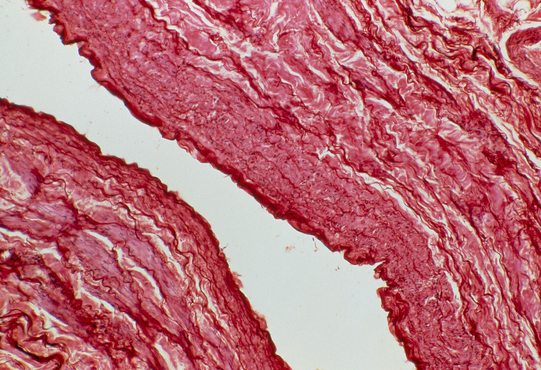
| Light micrograph of a longitudinal section of a large vein (lumen white). The thin inner layer of the wall (dark red),known as tunica intima,is formed by a flattened epithelium supported by a layer of connective tissue containing a few elastic fibres. The tunica intima is surrounded by the tunica media which consists of several layers of smooth muscle separated by layers of collagenous connective tissue. The last layer,the tunica adventitia is best seen at top right. In large veins the tunica adventitia is thicker than the tunica media and contains many longitudinal muscle fibres. Magnification: x25 at 35mm size | |
| Lizenzart: | Lizenzpflichtig |
| Credit: | Science Photo Library / Michler, Astrid & Hans-Frieder |
| Bildgröße: | 5056 px × 3461 px |
| Modell-Rechte: | nicht erforderlich |
| Eigentums-Rechte: | nicht erforderlich |
| Restrictions: | - |
Preise für dieses Bild ab 15 €
Universitäten & Organisationen
(Informationsmaterial Digital, Informationsmaterial Print, Lehrmaterial Digital etc.)
ab 15 €
Redaktionell
(Bücher, Bücher: Sach- und Fachliteratur, Digitale Medien (redaktionell) etc.)
ab 30 €
Werbung
(Anzeigen, Aussenwerbung, Digitale Medien, Fernsehwerbung, Karten, Werbemittel, Zeitschriften etc.)
ab 55 €
Handelsprodukte
(bedruckte Textilie, Kalender, Postkarte, Grußkarte, Verpackung etc.)
ab 75 €
Pauschalpreise
Rechtepakete für die unbeschränkte Bildnutzung in Print oder Online
ab 495 €