Cartilage cells
Bildnummer 11868514
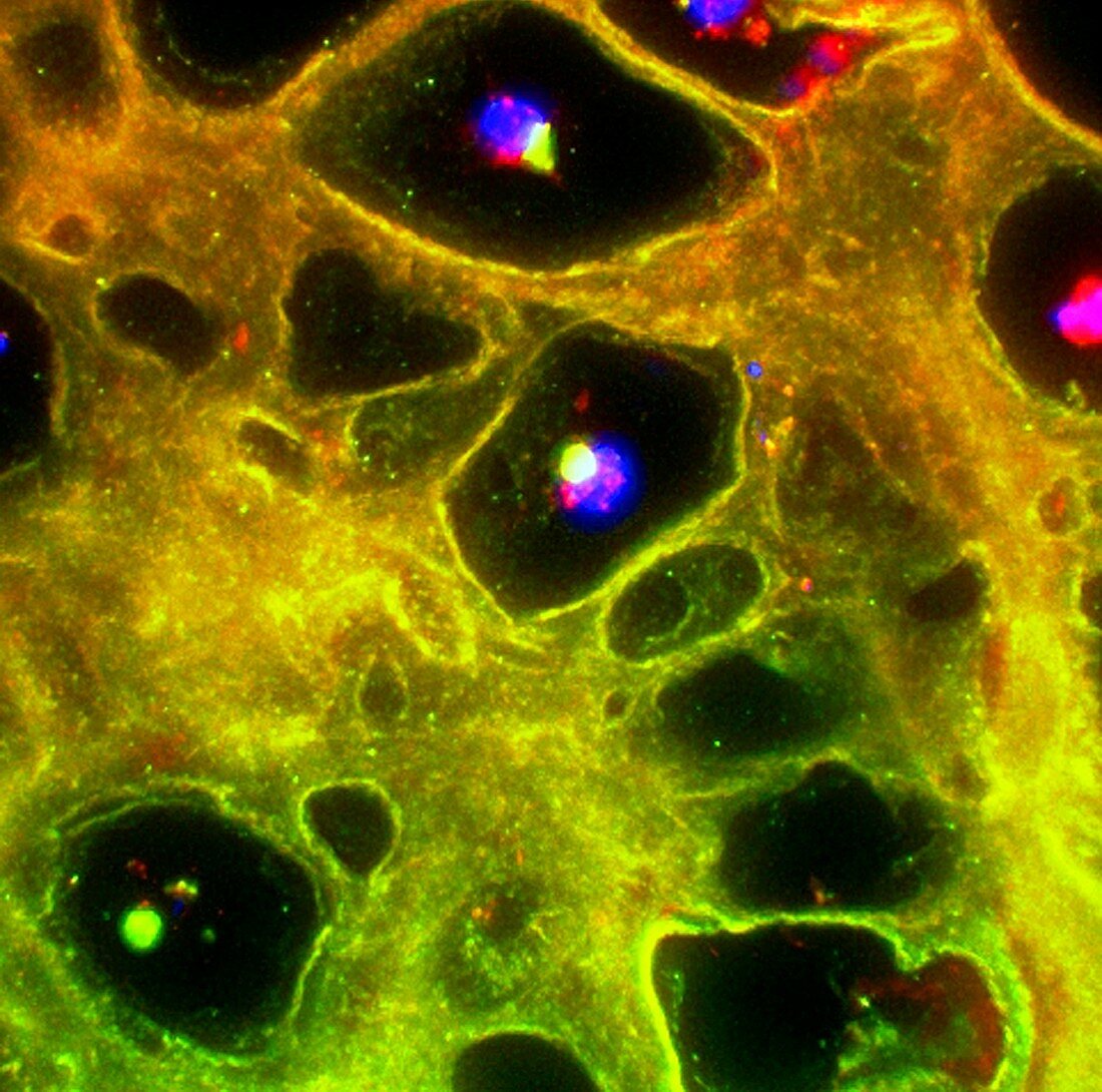
| Cartilage cells,fluorescence deconvolution micrograph. Fluorescent dyes have been used to highlight tissues and cellular structures: lacunae (black),cell nuclei (blue),cartilage matrix (orange,yellow and green). Lacunae are empty spaces,here in the matrix of cartilage (mainly collagen and elastin) secreted by cells known as chrondrocytes. The chrondrocyte nuclei are seen here | |
| Lizenzart: | Lizenzpflichtig |
| Credit: | Science Photo Library / R. BICK, B. POINDEXTER, UT MEDICAL SCHOOL |
| Bildgröße: | 3300 px × 3270 px |
| Modell-Rechte: | nicht erforderlich |
| Eigentums-Rechte: | nicht erforderlich |
| Restrictions: | - |
Preise für dieses Bild ab 15 €
Universitäten & Organisationen
(Informationsmaterial Digital, Informationsmaterial Print, Lehrmaterial Digital etc.)
ab 15 €
Redaktionell
(Bücher, Bücher: Sach- und Fachliteratur, Digitale Medien (redaktionell) etc.)
ab 30 €
Werbung
(Anzeigen, Aussenwerbung, Digitale Medien, Fernsehwerbung, Karten, Werbemittel, Zeitschriften etc.)
ab 55 €
Handelsprodukte
(bedruckte Textilie, Kalender, Postkarte, Grußkarte, Verpackung etc.)
ab 75 €
Pauschalpreise
Rechtepakete für die unbeschränkte Bildnutzung in Print oder Online
ab 495 €
Keywords
- Anatomie,
- anatomisch,
- Biochemie,
- biochemisch,
- dapi,
- Eiweiß,
- Farbstoff,
- Fluoreszenz,
- Fluoreszenzentfaltung,
- fluoreszierend,
- Gewebe,
- Histologie,
- histologisch,
- Knorpel,
- knorpelig,
- Knorpelmatrix,
- Lichtmikroskop,
- Marker,
- menschlicher Körper,
- Mikrofotografie,
- Zellbilogie,
- Zelle,
- Zellen,
- Zellkern,
- Zytologie