Child's head and chest,CT scan
Bildnummer 11868148
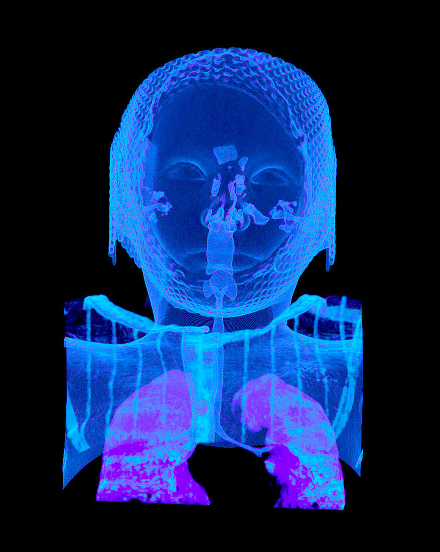
| Child's head and chest,CT scan. Coloured 3-D computed tomography (CT) scan of a child's head and chest in frontal view. Within the head,the scan shows the sinuses,ear passages and nasal cavity. Within the throat and chest,the scan shows the larynx,pharynx,trachea and lungs. The scan will be used by surgeons to navigate around the brain while operating for brain cancer. This image was produced using a multi-slice CT scanner,which uses a thin X-ray beam to scan around the patient. OsiriX medical imaging software was used to reconstruct the slices into coloured 3-D images of bones and soft tissue. The program allows surgeons to navigate around the body using fly- through animations | |
| Lizenzart: | Lizenzpflichtig |
| Credit: | Science Photo Library / Rosset, Antoine |
| Bildgröße: | 2647 px × 3319 px |
| Modell-Rechte: | nicht erforderlich |
| Eigentums-Rechte: | nicht erforderlich |
| Restrictions: | - |
Preise für dieses Bild ab 15 €
Universitäten & Organisationen
(Informationsmaterial Digital, Informationsmaterial Print, Lehrmaterial Digital etc.)
ab 15 €
Redaktionell
(Bücher, Bücher: Sach- und Fachliteratur, Digitale Medien (redaktionell) etc.)
ab 30 €
Werbung
(Anzeigen, Aussenwerbung, Digitale Medien, Fernsehwerbung, Karten, Werbemittel, Zeitschriften etc.)
ab 55 €
Handelsprodukte
(bedruckte Textilie, Kalender, Postkarte, Grußkarte, Verpackung etc.)
ab 75 €
Pauschalpreise
Rechtepakete für die unbeschränkte Bildnutzung in Print oder Online
ab 495 €
Keywords
- 3-d,
- 3D,
- Anatomie,
- anatomisch,
- Angiogramm,
- Arterien,
- ärztliche Untersuchung,
- Bronchien,
- Computertomographie,
- CT-Scan,
- CTA,
- Diagnose,
- diagnostische Bildgebung,
- Dreidimensional,
- fazial,
- Halsschlagader,
- Kind,
- Knochen,
- Kontrastmittel,
- Kopf,
- Lungen,
- Medizin,
- medizinisch,
- medizinische Bildgebung,
- medizinische Visualisierung,
- medizinischer Scan,
- Mensch,
- Menschen,
- menschlicher Körper,
- Nebenhöhlen,
- OsiriX,
- Person,
- Radiographie,
- Radiologie,
- radiologisch,
- Röntgen,
- Röntgenstrahlen,
- Röntgenstrahlung,
- Scanner,
- Schädel,
- Sinus,
- Truhe