3-D CT scan of human skull
Bildnummer 11868072
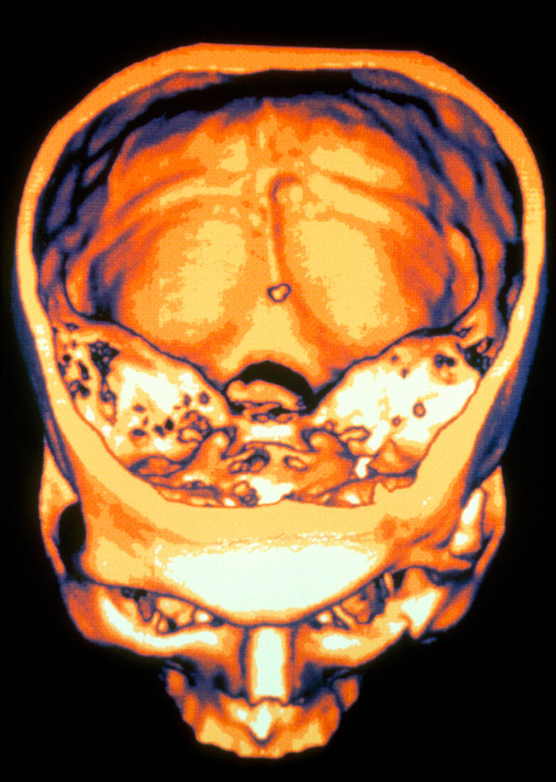
| Human skull. Three-dimensional computed tomography (CT) scan of the human skull seen from above,with the top of the cranium removed. The cranium chamber (at upper centre) houses the brain. At centre is the foramen magnum,the hole in base of the skull through which the spinal cord passes. The petrous portion of the temporal bone (at far left and right) contains the petrosal sinus (at centre). The orbits (eye sockets),nasal bones and maxillae (upper jaw) are at lower centre. Zygomatic arches (at lower left and right) form the cheek bones. CT scans use X-rays to form "slices" of body tissue. These are built up into a three-dimensional image by computer | |
| Lizenzart: | Lizenzpflichtig |
| Credit: | Science Photo Library / GCA |
| Bildgröße: | 2518 px × 3543 px |
| Modell-Rechte: | nicht erforderlich |
| Eigentums-Rechte: | nicht erforderlich |
| Restrictions: | - |
Preise für dieses Bild ab 15 €
Universitäten & Organisationen
(Informationsmaterial Digital, Informationsmaterial Print, Lehrmaterial Digital etc.)
ab 15 €
Redaktionell
(Bücher, Bücher: Sach- und Fachliteratur, Digitale Medien (redaktionell) etc.)
ab 30 €
Werbung
(Anzeigen, Aussenwerbung, Digitale Medien, Fernsehwerbung, Karten, Werbemittel, Zeitschriften etc.)
ab 55 €
Handelsprodukte
(bedruckte Textilie, Kalender, Postkarte, Grußkarte, Verpackung etc.)
ab 75 €
Pauschalpreise
Rechtepakete für die unbeschränkte Bildnutzung in Print oder Online
ab 495 €