False-colour bone scintigram of a human skull
Bildnummer 11868035
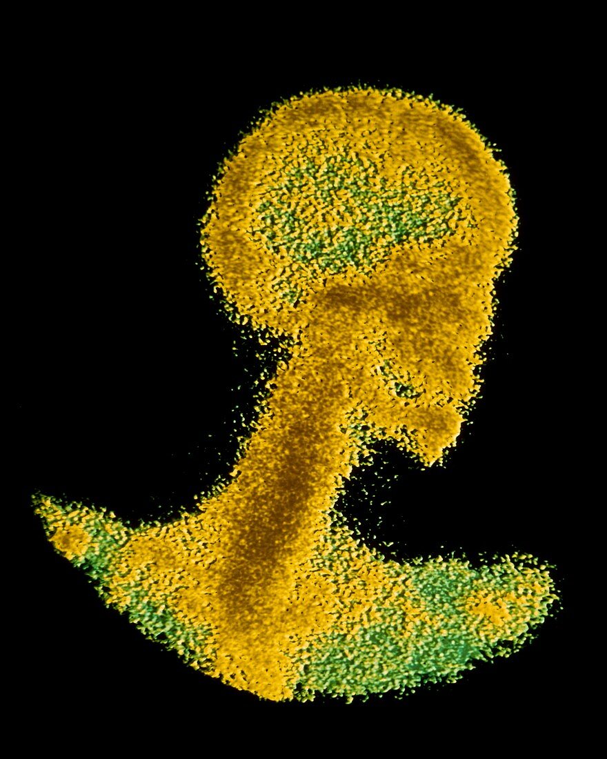
| False-colour bone scintigram of the head,neck & skull areas of a normal,healthy person. The image is produced by recording the distribution of gamma rays emitted after a small amount of a radio- isotope has been injected into the patient. The gamma rays are recorded by the light they give off when they strike a scintillating material in a "gamma camera". In this instance the radiation uptake in bone is being measured,a procedure which is typically used to assess the presence or extent of secondary bone cancers. Cancerous bone absorbs the radio-isotope more strongly than healthy tissue,& produces a brighter,"hotter" area on the image | |
| Lizenzart: | Lizenzpflichtig |
| Credit: | Science Photo Library / Pol, Alain / ISM |
| Bildgröße: | 3780 px × 4724 px |
| Modell-Rechte: | nicht erforderlich |
| Eigentums-Rechte: | nicht erforderlich |
| Restrictions: |
|
Preise für dieses Bild ab 15 €
Universitäten & Organisationen
(Informationsmaterial Digital, Informationsmaterial Print, Lehrmaterial Digital etc.)
ab 15 €
Redaktionell
(Bücher, Bücher: Sach- und Fachliteratur, Digitale Medien (redaktionell) etc.)
ab 30 €
Werbung
(Anzeigen, Aussenwerbung, Digitale Medien, Fernsehwerbung, Karten, Werbemittel, Zeitschriften etc.)
ab 55 €
Handelsprodukte
(bedruckte Textilie, Kalender, Postkarte, Grußkarte, Verpackung etc.)
ab 75 €
Pauschalpreise
Rechtepakete für die unbeschränkte Bildnutzung in Print oder Online
ab 495 €