Female pelvis,coloured 3-D CT scan
Bildnummer 11867875
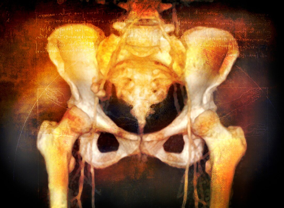
| Female pelvis,coloured 3-D computed tomography (CT) scan. The upper part of each thigh bone (femur) is seen at each side of the pelvis,where they form the hip joints. The spine runs down centre,terminating in the coccyx (tail bone) at lower centre. Major blood vessels are also seen,separating at top centre and running down each leg. The female pelvis is wider and shallower than the male,which facilitates childbirth | |
| Lizenzart: | Lizenzpflichtig |
| Credit: | Science Photo Library / Maslo, Miriam |
| Bildgröße: | 4882 px × 3580 px |
| Modell-Rechte: | nicht erforderlich |
| Eigentums-Rechte: | nicht erforderlich |
| Restrictions: | - |
Preise für dieses Bild ab 15 €
Universitäten & Organisationen
(Informationsmaterial Digital, Informationsmaterial Print, Lehrmaterial Digital etc.)
ab 15 €
Redaktionell
(Bücher, Bücher: Sach- und Fachliteratur, Digitale Medien (redaktionell) etc.)
ab 30 €
Werbung
(Anzeigen, Aussenwerbung, Digitale Medien, Fernsehwerbung, Karten, Werbemittel, Zeitschriften etc.)
ab 55 €
Handelsprodukte
(bedruckte Textilie, Kalender, Postkarte, Grußkarte, Verpackung etc.)
ab 75 €
Pauschalpreise
Rechtepakete für die unbeschränkte Bildnutzung in Print oder Online
ab 495 €
Keywords
- Anatomie,
- Becken,
- Blutgefäß,
- Blutgefäße,
- Computertomographie,
- CT-Scan,
- Diagnose,
- Dreidimensional,
- einer,
- Femur,
- Frau,
- Hüfte,
- Hüften,
- Illustration,
- Knochen,
- Medizin,
- medizinisch,
- menschlicher Körper,
- Orange,
- Physiologie,
- physiologisch,
- Radiographie,
- Scanner,
- Single,
- Skelett,
- Skelett-,
- Weiblich,
- Wirbelsäule,
- Wirbelsäulen-