Lower spine,bone density scan
Bildnummer 11867820
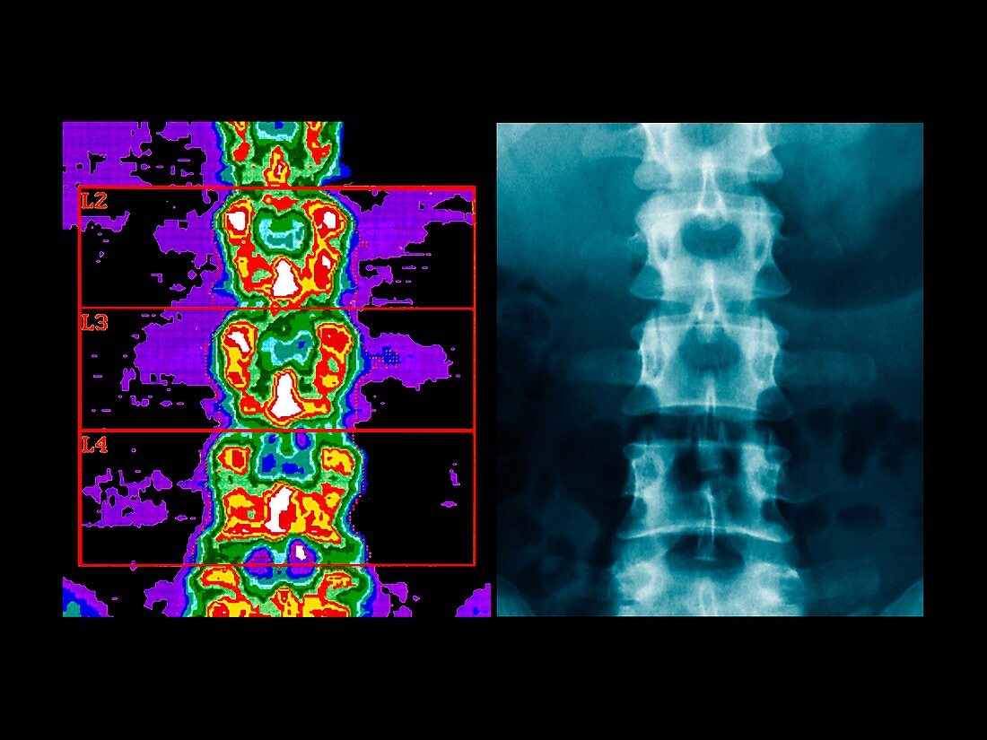
| Spinal scan and X-ray. Coloured bone densitometry scan (left) and an X-ray (right),of the same area of the spine. This is a frontal view of the lumbar (lower) spine. From top,the vertebrae L2,L3,and L4 are marked. A bone densitometry scan uses X- rays to measure bone density. The bone density is colour-coded,ranging from blue/purple (least dense) through to white (most dense). The average bone density of this menopausal patient is normal. The scan is often used to detect decreased bone density (osteoporosis). The vertebrae are the blocks of bone that make up the spine. There are 24 vertebrae making up the flexible part of the spine,of which 5 are in the lumbar region | |
| Lizenzart: | Lizenzpflichtig |
| Credit: | Science Photo Library / Zephyr |
| Bildgröße: | 4724 px × 3543 px |
| Modell-Rechte: | nicht erforderlich |
| Eigentums-Rechte: | nicht erforderlich |
| Restrictions: | - |
Preise für dieses Bild ab 15 €
Universitäten & Organisationen
(Informationsmaterial Digital, Informationsmaterial Print, Lehrmaterial Digital etc.)
ab 15 €
Redaktionell
(Bücher, Bücher: Sach- und Fachliteratur, Digitale Medien (redaktionell) etc.)
ab 30 €
Werbung
(Anzeigen, Aussenwerbung, Digitale Medien, Fernsehwerbung, Karten, Werbemittel, Zeitschriften etc.)
ab 55 €
Handelsprodukte
(bedruckte Textilie, Kalender, Postkarte, Grußkarte, Verpackung etc.)
ab 75 €
Pauschalpreise
Rechtepakete für die unbeschränkte Bildnutzung in Print oder Online
ab 495 €
Keywords
- Anatomie,
- Densitometrie,
- Diagnose,
- farbig,
- Frau,
- Frontal,
- geduldig,
- Knochen,
- L2,
- L3,
- L4,
- Lendenwirbelsäule,
- menschlicher Körper,
- Osteologie,
- osteologisch,
- Radiographie,
- Radiologie,
- Röntgen,
- Röntgenbild,
- Rückgrat,
- Sektion,
- sektioniert,
- Skelett,
- Stirnbein,
- unterer Rücken,
- Vergleich,
- vergleichen,
- Weiblich,
- Wirbel,
- Wirbelsäule,
- Wirbelsäulen-