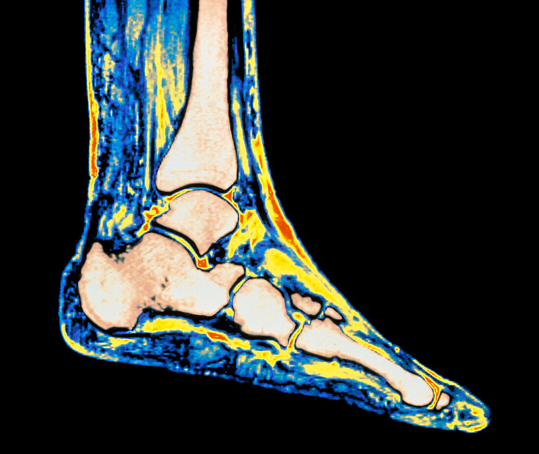
Coloured MRI scan of ankle bones in the human foot
Bildnummer 11867690

| Bones of the ankle. Magnetic Resonance Imaging (MRI) scan of a sagittal (side) view through the healthy ankle of a woman aged 58. The ankle forms a hinge joint between the leg (at top) and foot (lower frame). Bones are beige coloured; soft tissue is brightly coloured. The tibia is the main leg bone seen; its concave head articulates with the cube-shaped talus,the uppermost bone in the foot. The talus is above the calcaneus bone which forms the heel projection behind the foot. The talus and calcaneus attach to tarsal bones in the middle of the foot,which in turn articulate with long metatarsal bones at the base of the toes | |
| Lizenzart: | Lizenzpflichtig |
| Credit: | Science Photo Library / Fraser, Simon |
| Bildgröße: | 4543 px × 3823 px |
| Modell-Rechte: | nicht erforderlich |
| Eigentums-Rechte: | nicht erforderlich |
| Restrictions: | - |
Preise für dieses Bild ab 15 €
Universitäten & Organisationen
(Informationsmaterial Digital, Informationsmaterial Print, Lehrmaterial Digital etc.)
ab 15 €
Redaktionell
(Bücher, Bücher: Sach- und Fachliteratur, Digitale Medien (redaktionell) etc.)
ab 30 €
Werbung
(Anzeigen, Aussenwerbung, Digitale Medien, Fernsehwerbung, Karten, Werbemittel, Zeitschriften etc.)
ab 55 €
Handelsprodukte
(bedruckte Textilie, Kalender, Postkarte, Grußkarte, Verpackung etc.)
ab 75 €
Pauschalpreise
Rechtepakete für die unbeschränkte Bildnutzung in Print oder Online
ab 495 €
