3-D CT scan of bones of a normal adult pelvis
Bildnummer 11867675
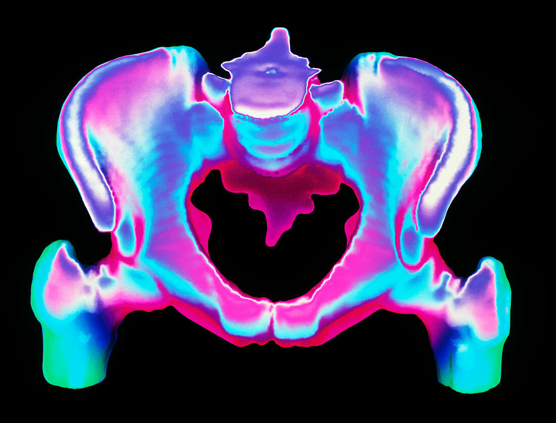
| Human pelvis. Coloured three-dimensional Computed Tomography (CT) scan of bones of a normal adult pelvis. At top centre is the flattened region where lumbar vertebrae articulate with the pelvis; at lower left & right is the rounded head of each femur (thigh bone) which fit into hip sockets of the pelvis. The pelvis itself is formed by bones of the sacrum and coccyx (fused vertebrae,at centre) and two curved innominate or hip bones. The pelvis supports and protects internal organs and tissues of the abdomen,and provides a site for the attachment of muscles of the trunk and lower limbs | |
| Lizenzart: | Lizenzpflichtig |
| Credit: | Science Photo Library |
| Bildgröße: | 4961 px × 3776 px |
| Modell-Rechte: | nicht erforderlich |
| Eigentums-Rechte: | nicht erforderlich |
| Restrictions: | - |
Preise für dieses Bild ab 15 €
Universitäten & Organisationen
(Informationsmaterial Digital, Informationsmaterial Print, Lehrmaterial Digital etc.)
ab 15 €
Redaktionell
(Bücher, Bücher: Sach- und Fachliteratur, Digitale Medien (redaktionell) etc.)
ab 30 €
Werbung
(Anzeigen, Aussenwerbung, Digitale Medien, Fernsehwerbung, Karten, Werbemittel, Zeitschriften etc.)
ab 55 €
Handelsprodukte
(bedruckte Textilie, Kalender, Postkarte, Grußkarte, Verpackung etc.)
ab 75 €
Pauschalpreise
Rechtepakete für die unbeschränkte Bildnutzung in Print oder Online
ab 495 €