Lumbar myelogram showing residual contrast oil
Bildnummer 11867625
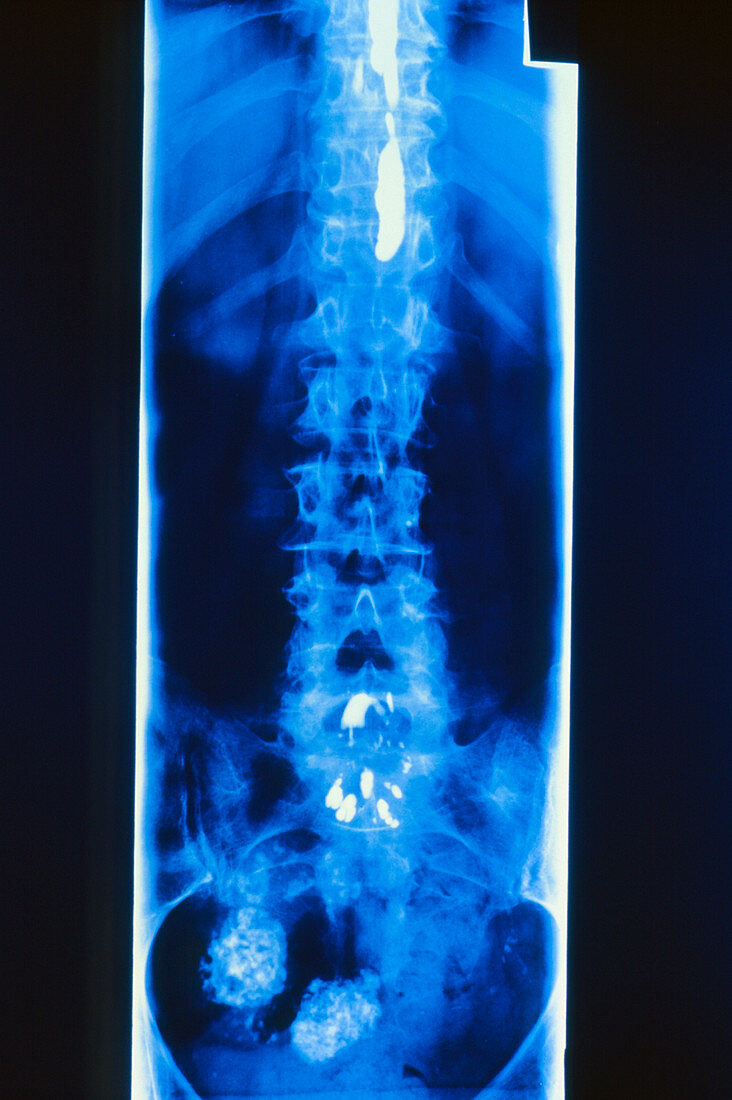
| After effects of myelography. Ordinary X-ray image of the human spine taken two years after the subject had undergone myelography examination,showing residual oily contrast material (myodil) in the spinal canal (white areas). Myelography is used to examine the neural tissue within the spine through the introduction of a radio-opaque contrast medium (iodized water or oil-based) into the sub-arachnoid space at lumbar puncture. The present material of choice is water-based,due to the frequency of complications from arachnoiditis associated with iodized oil such as myodil. Myelography confined to the lumbar spine is termed radiculography | |
| Lizenzart: | Lizenzpflichtig |
| Credit: | Science Photo Library |
| Bildgröße: | 2480 px × 3726 px |
| Modell-Rechte: | nicht erforderlich |
| Eigentums-Rechte: | nicht erforderlich |
| Restrictions: | - |
Preise für dieses Bild ab 15 €
Universitäten & Organisationen
(Informationsmaterial Digital, Informationsmaterial Print, Lehrmaterial Digital etc.)
ab 15 €
Redaktionell
(Bücher, Bücher: Sach- und Fachliteratur, Digitale Medien (redaktionell) etc.)
ab 30 €
Werbung
(Anzeigen, Aussenwerbung, Digitale Medien, Fernsehwerbung, Karten, Werbemittel, Zeitschriften etc.)
ab 55 €
Handelsprodukte
(bedruckte Textilie, Kalender, Postkarte, Grußkarte, Verpackung etc.)
ab 75 €
Pauschalpreise
Rechtepakete für die unbeschränkte Bildnutzung in Print oder Online
ab 495 €