Human foetus
Bildnummer 11867078
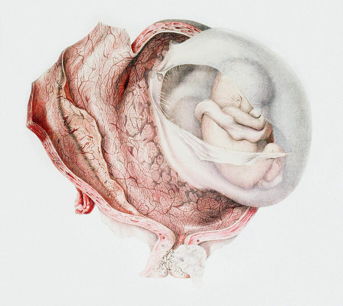
| Human foetus. Historical anatomical artwork of a human foetus in the uterus. The uterus has been sliced open to reveal the foetus in its chorionic sac membrane,which has also been sliced open. The umbilical cord is visible,as are the network of blood vessels on the inner surface of the uterus. The cervix,the neck of the uterus,is at lower centre. This foetus is in the third to fourth month of gestation. Artwork from the 19th-century book Atlas of Anatomy,by Bourgery and Jacob. This book,which took over 20 years to complete,was published in France in 8 volumes from 1831 to 1854. It contained 726 colour plates covering both anatomy and surgical techniques | |
| Lizenzart: | Lizenzpflichtig |
| Credit: | Science Photo Library / Kulyk, Mehau |
| Bildgröße: | 4550 px × 4068 px |
| Modell-Rechte: | nicht erforderlich |
| Eigentums-Rechte: | nicht erforderlich |
| Restrictions: | - |
Preise für dieses Bild ab 15 €
Universitäten & Organisationen
(Informationsmaterial Digital, Informationsmaterial Print, Lehrmaterial Digital etc.)
ab 15 €
Redaktionell
(Bücher, Bücher: Sach- und Fachliteratur, Digitale Medien (redaktionell) etc.)
ab 30 €
Werbung
(Anzeigen, Aussenwerbung, Digitale Medien, Fernsehwerbung, Karten, Werbemittel, Zeitschriften etc.)
ab 55 €
Handelsprodukte
(bedruckte Textilie, Kalender, Postkarte, Grußkarte, Verpackung etc.)
ab 75 €
Pauschalpreise
Rechtepakete für die unbeschränkte Bildnutzung in Print oder Online
ab 495 €
Keywords
- 1800er Jahre,
- 19. Jahrhundert,
- Abdomen,
- Anatomie,
- anatomisch,
- aufgegliedert,
- Biologie,
- biologisch,
- Erwachsene,
- Fortpflanzungssystem,
- Fötus,
- Französisch,
- Frau,
- Geburtshilfe,
- Geschichte,
- historisch,
- Illustration,
- Jean Baptiste Marc Bourgery,
- kaukasisch,
- Leiche,
- medizinisch,
- Mensch,
- menschlicher Körper,
- Nicolas Henri Jacob,
- pränatal,
- Reproduktion,
- schwanger,
- Schwangerschaft,
- uterin,
- Uterus,
- Weiblich,
- weiß,
- Zeichnung