Embryonic development
Bildnummer 11866590
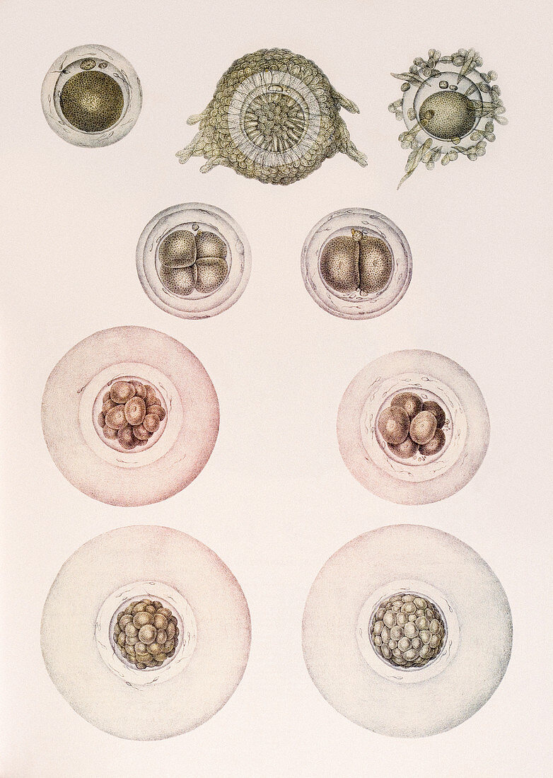
| Development of an embryo,historical anatomical artwork. This 19th century textbook illustration shows the sequence of stages in embryonic development,starting at upper left. The first diagram is an ovum (egg cell) removed from the ovary of a rabbit. The second and third show the process of fertilisation with the ovum surrounded by sperm cells. Once fertilised,the cell divides (central diagrams). The final diagram shows a blastocyst,or hollow ball of cells. Development takes place in the fallopian tube,as the cells travel towards the uterus. The illustration is taken from the 19th century French textbook The Atlas of Human Anatomy and Surgery by J. M. Bourgery and N. H. Jacob | |
| Lizenzart: | Lizenzpflichtig |
| Credit: | Science Photo Library / Kulyk, Mehau |
| Bildgröße: | 3822 px × 5376 px |
| Modell-Rechte: | nicht erforderlich |
| Eigentums-Rechte: | nicht erforderlich |
| Restrictions: | - |
Preise für dieses Bild ab 15 €
Universitäten & Organisationen
(Informationsmaterial Digital, Informationsmaterial Print, Lehrmaterial Digital etc.)
ab 15 €
Redaktionell
(Bücher, Bücher: Sach- und Fachliteratur, Digitale Medien (redaktionell) etc.)
ab 30 €
Werbung
(Anzeigen, Aussenwerbung, Digitale Medien, Fernsehwerbung, Karten, Werbemittel, Zeitschriften etc.)
ab 55 €
Handelsprodukte
(bedruckte Textilie, Kalender, Postkarte, Grußkarte, Verpackung etc.)
ab 75 €
Pauschalpreise
Rechtepakete für die unbeschränkte Bildnutzung in Print oder Online
ab 495 €
Keywords
- 1800er Jahre,
- 19. Jahrhundert,
- Anatomie,
- anatomisch,
- Bilderwelt,
- Biologie,
- biologisch,
- Blastomere,
- Blastozyste,
- chirurgisch,
- Einteilung,
- Embryologie,
- Entwicklung,
- entwicklungsgemäß,
- Französisch,
- Geschichte,
- historisch,
- historisches Bild,
- Illustration,
- Kunstwerk,
- mehrere,
- menschlicher Körper,
- Morula,
- Operation,
- Reproduktion,
- reproduktiv,
- Sperma,
- Spermatozoen,
- Teilen,
- viele,
- Zelle,
- Zygote